
Blood cells turned directly into neural stem cells read as epigenetically young — in lab-grown cells
Edited by Samael A. Tejada, Founder & Editor-in-Chief
·Tip: press play, then lock your phone or switch apps — the article keeps reading.
Epigenetic clocks are built largely from methylation marks on nuclear DNA, and cellular youth is bigger than any one clock. What Bonn showed is narrower and still remarkable: a direct-conversion route that skips the pluripotent stage and lands on neural stem cells whose methylation age reads decades younger than the person they came from. Whether that younger reading means younger mitochondria, younger metabolism or a younger cell in any functional sense is exactly what this study did not test.
What exactly did the Bonn team do?
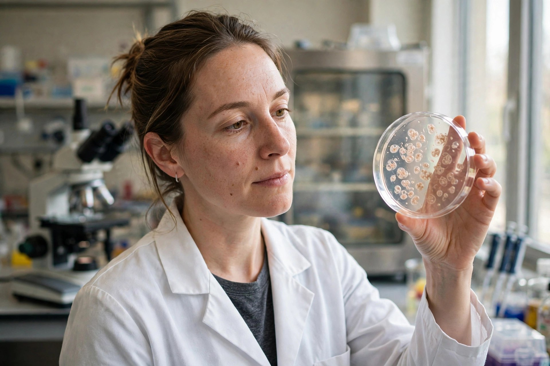
The method starts with peripheral blood mononuclear cells — the mixed immune-cell fraction from a blood sample — taken from 15 donors spanning newborn to 101 years old. Instead of first pushing those cells back to induced pluripotent stem cells (the standard route, with its extra time, cost and tumor-risk questions), the team converted them directly into induced neural stem cells. The epigenetic reset was not instant: methylation age declined gradually across about 50 days of conversion until the resulting neural stem cells read as young adults on the clock, at less than 20 years of epigenetic age regardless of donor age.
How large was the age shift, in plain terms?
For an 80-year-old donor, the converted cells' epigenetic age fell by more than 60 years — landing at roughly 5 percent of the donor's chronological age. The same young endpoint was reached across the donor panel, which is why the result drew coverage beyond the stem-cell field: it suggests the conversion process itself, not the donor, sets the epigenetic age of the product. The authors' identified uses are laboratory ones — disease modeling and research into brain aging — where young-reading neural stem cells from an old donor are a tool for asking what aging changed, not a therapy for changing it back in a person.
What does this mean for mitochondria — and what does it not mean?
Readers of this site will ask the mitochondrial question, so here is the precise answer. Epigenetic clocks are calculated from CpG methylation sites that sit overwhelmingly outside the mitochondrial genome, and this paper reports no mitochondrial respiration or mitochondrial DNA measures among its findings; its one mitochondrial assay, a reactive-oxygen-species hallmark check in established cell lines, read comparable to young-like control cells. Aging biology gives reasons to suspect mitochondrial state and epigenetic state move together, and mitochondria do supply metabolites the methylation machinery uses; but suspicion is not measurement. The mitochondrial claim for this story is therefore absent, and the follow-up experiment that would change that — respiration and mtDNA profiling of the converted cells against donor-matched controls — has not been reported.
Frequently asked questions
What did the Bonn researchers make from blood cells?
Induced neural stem cells, made by direct conversion of peripheral blood mononuclear cells with no pluripotent stem-cell stage in between, from 15 donors ranging from newborns to a 101-year-old.
How young did the converted cells read?
On DNA methylation (epigenetic) clocks, the neural stem cells read as under 20 years old after about 50 days of conversion — roughly 5 percent of the donor's age, and a shift of more than 60 years for cells from an 80-year-old donor.
Is this a treatment that reverses aging in people?
No. The cells were grown and measured in a laboratory dish. No animal or person received them, and safety, stability and brain function are unproven. The study also did not measure mitochondria, so it makes no mitochondrial claim.
Where the evidence stands
- Established: Direct lineage conversion can turn accessible blood cells into neural stem cells without a pluripotent intermediate, and epigenetic (DNA methylation) clocks can estimate a cell population's biological age independently of the donor's chronological age.
- Preliminary: In lab-grown cells from 15 human donors aged newborn to 101 (Aging Cell, 2026; DOI 10.1111/acel.70751), direct conversion produced neural stem cells whose epigenetic age fell gradually over about 50 days to under 20 years — about 5 percent of donor age, a shift of more than 60 years for an 80-year-old donor.
- Absent: Any in-vivo or clinical result, any safety or tumor-risk data in a living organism, any demonstration of function in a brain, and any mitochondrial respiration, mtDNA or dynamics measurement in the converted cells (the one mitochondrial assay reported, a ROS hallmark check in established lines, read comparable to young-like control cells).
Join the discussion
To get the conversation started: What questions does this story raise for you? Share them below — every comment is reviewed by our moderators before it appears.
Community discussion only — not medical advice. Every comment is reviewed by our moderators before it appears. Sign in with Google or a free Hyvor account to join in.