
Research
New findings from the laboratory and clinic, labeled by evidence maturity.

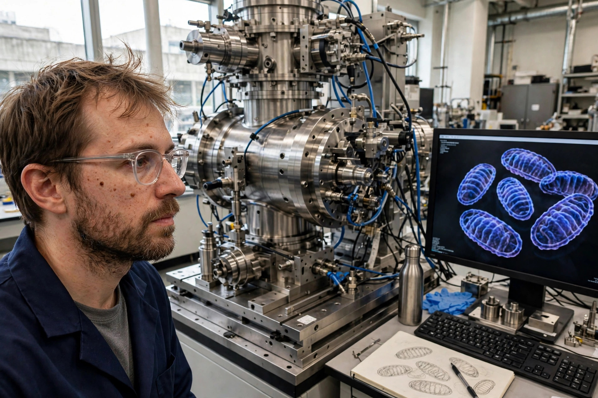
Sharper math, not sharper lenses: X-ray imaging can now pick out a single mitochondrion in 3D
Seeing a mitochondrion inside an intact sample, in three dimensions, has usually meant electron microscopy — which requires cutting material into thin slices and damages it. An international team reported in Optica (Nikitin, Carlsson and colleagues; covered November 10, 2026) that a new mathematical reconstruction method for X-ray nano-holotomography improves image resolution about threefold, distinguishing structures down to 20 nanometers — a human hair is 80,000-100,000 nanometers thick. At that resolution, synapses, mitochondria and other subcellular structures become visible in 3D in much larger samples, without slicing them apart. Last author Marcus Carlsson of Lund University puts the point plainly: the limitation had been not only the X-ray optics, but the mathematics used to reconstruct the image.
Read article →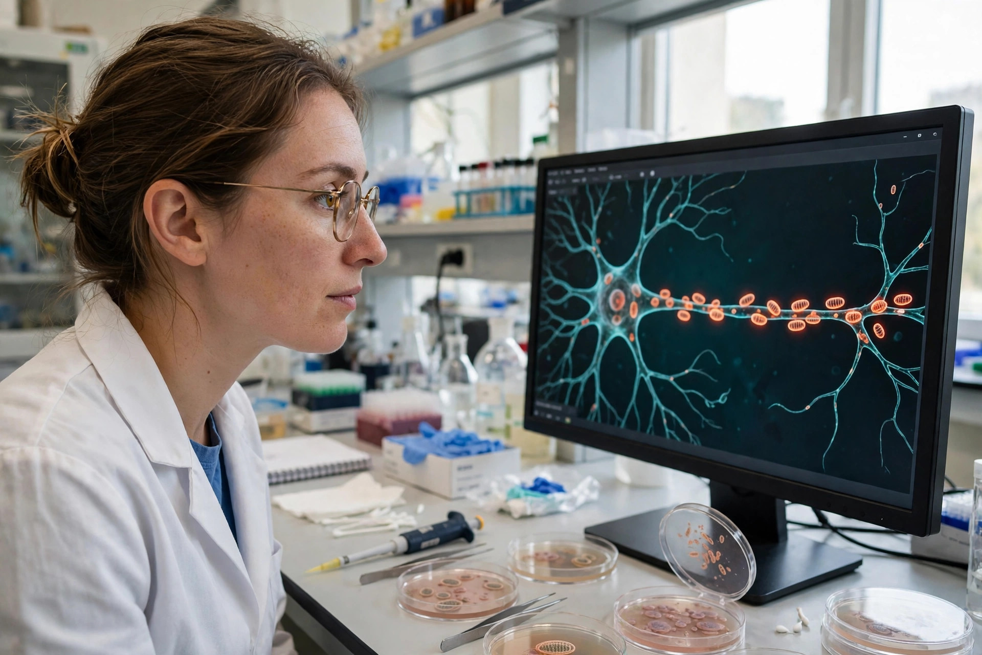
A contact site acts as a loading dock for mitochondria moving down nerve cells — in mice and cells
Neurons are long, and their mitochondria must be shipped down axons to where energy is needed. A study in Cell Discovery (Pei, Zhang, Li and colleagues; published November 10, 2026) identifies C19orf12 — a protein mutated in spastic paraplegia type 43 (SPG43) and in a form of neurodegeneration with brain iron accumulation — as an endoplasmic-reticulum-anchored organizer of mitochondrial transport, enriched where the ER touches mitochondria. The authors propose renaming it EMTRAS (ER-anchored Mitochondrial Trafficking Scaffold). C19orf12 deficiency drove progressive motor impairment in mice and distal mitochondrial depletion in neurons; mechanistically, it works as a calcium-tunable scaffold that promotes assembly of the Miro1-Kinesin-1 transport complex, linking ER-mitochondria contact sites directly to microtubule-based movement.
Read article →
Tiny packets shed by mitochondria may help spread Alzheimer's damage — in human tissue and mice
Cells release tiny membrane-wrapped packets, and one subset — mitovesicles — comes from mitochondria. A study in Nature Aging (Li, Xia, Zheng and colleagues; published November 10, 2026) found that release of mitochondria-derived vesicles is increased in the brains of Alzheimer's patients and of 5xFAD model mice, and that these vesicles are enriched for the enzyme Ggt1, which breaks down the antioxidant glutathione. Injecting vesicles from 5xFAD brains into healthy mice impaired learning and memory and drove glial inflammation and synaptic abnormalities, and accelerated disease in Alzheimer's-model mice. Knocking out or pharmacologically inhibiting Ggt1 mitigated those effects, and suppressing vesicle release with fumarate-hydratase inhibition or dimethyl fumarate/coenzyme Q10 also alleviated the phenotypes in the animal model.
Read article →
When mitochondria fail, the brain's protective barrier may fail with them — a review of the evidence
The blood-brain barrier decides what reaches the brain, and its cells are among the most mitochondria-dependent in the body. A review in Fluids and Barriers of the CNS (Cicala, Geldenhuys, Huber and colleagues; published November 10, 2026) examines the mechanistic connection between mitochondrial dysfunction and breakdown of that barrier, with particular attention to how mitochondrial reactive oxygen species contribute to barrier dysfunction. The review synthesizes existing evidence linking mitochondrial health to barrier integrity and frames mitochondria as a promising target for cerebrovascular disorders. A review is a map of other people's experiments, not a new experiment: it can show where the evidence converges, and it cannot by itself prove that fixing mitochondria would protect the barrier in patients.
Read article →
The folds inside mitochondria may throttle a skin drug's inflammation — in a mouse-cell preprint
Imiquimod, an approved drug for skin malignancies, frequently provokes severe inflammation by activating the NLRP3 inflammasome, and its inhibition of mitochondrial complex I is necessary for that activation but not sufficient — leaving the second trigger in dispute. A bioRxiv preprint from a UCLA team (Srikanth, Wu, Gwack and colleagues; posted November 6, 2026; not peer-reviewed) used macrophages lacking TMEM11, a MICOS-associated inner-membrane protein that helps organize cristae — the folded membranes inside mitochondria. Tmem11-deficient macrophages showed ultrastructural cristae defects without altered mitochondrial mass or mtDNA copy number, responded normally to canonical inflammasome agonists (ATP, nigericin), but showed caspase-1 hyperactivation and exaggerated IL-1beta and IL-18 secretion on imiquimod treatment. The authors conclude cristae architecture restrains drug-induced mitochondrial reactive oxygen species, and that mtROS is a primary driver of imiquimod-mediated NLRP3 activation. Because this is a preprint, every claim here awaits peer review.
Read article →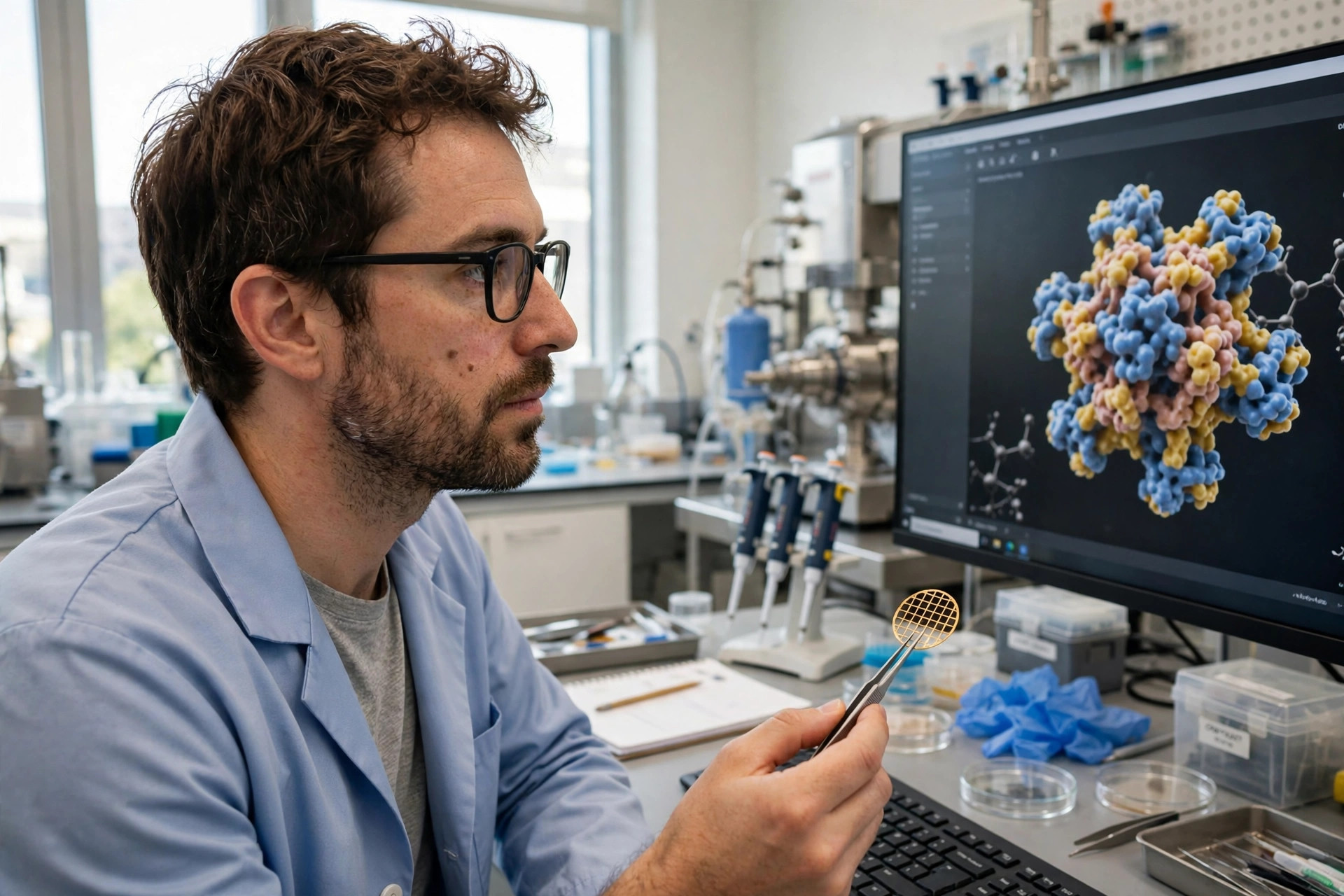
How mitochondria keep protein production moving: a helper protein caught in the act — in lab models
Mitochondria make a small but essential set of proteins on their own ribosomes (mitoribosomes), and some amino-acid sequences — notably runs of proline — make the mitoribosome slow or stall. A Karolinska Institutet study in Nature Communications (Singh, Rorbach, Khawaja and colleagues; reported November 5, 2026) captured human TACO1, a factor known to help the mitoribosome through such difficult sequences, in the act: using cryo-electron microscopy, the team imaged TACO1 bound to a mitoribosome mid-production, finding that it sits close to the ribosome's A site and interacts directly with the incoming transfer RNA, with large movements that help stabilize that tRNA. TACO1 works differently from related factors in bacteria and elsewhere in human cells, though it shares structural similarities with its bacterial relative YebC2. Mutations in TACO1 are associated with cytochrome c oxidase deficiency and late-onset Leigh syndrome; the structure offers a molecular explanation for how those changes interfere with mitochondrial protein synthesis.
Read article →
A redox enzyme protected lens cells' mitochondria in diabetic cataract — in mice and cells
In diabetic cataract, high glucose damages the mitochondria of lens epithelial cells, and that damage helps drive the disease. A study in Cell Biology and Toxicology (Zhang, Liang, Guo and colleagues; published November 9, 2026) found that glutaredoxin 2 (Grx2) protects those cells by activating the AMPK/PGC1alpha/NRF1 signaling pathway, which preserves PACS2-dependent integrity of mitochondria-associated endoplasmic reticulum membranes (MAMs) — the contact sites where mitochondria and the endoplasmic reticulum meet — and maintains mitochondrial homeostasis. The team used streptozotocin-induced diabetic mice with Grx2 knocked in or knocked out, lens tissue from those animals, and cultured HLE-B3 human lens epithelial cells under high glucose. Knocking Grx2 out aggravated protein glutathionylation (PSSG) accumulation, MAM disruption and mitochondrial dysfunction in diabetic mice; knocking it in rescued those defects.
Read article →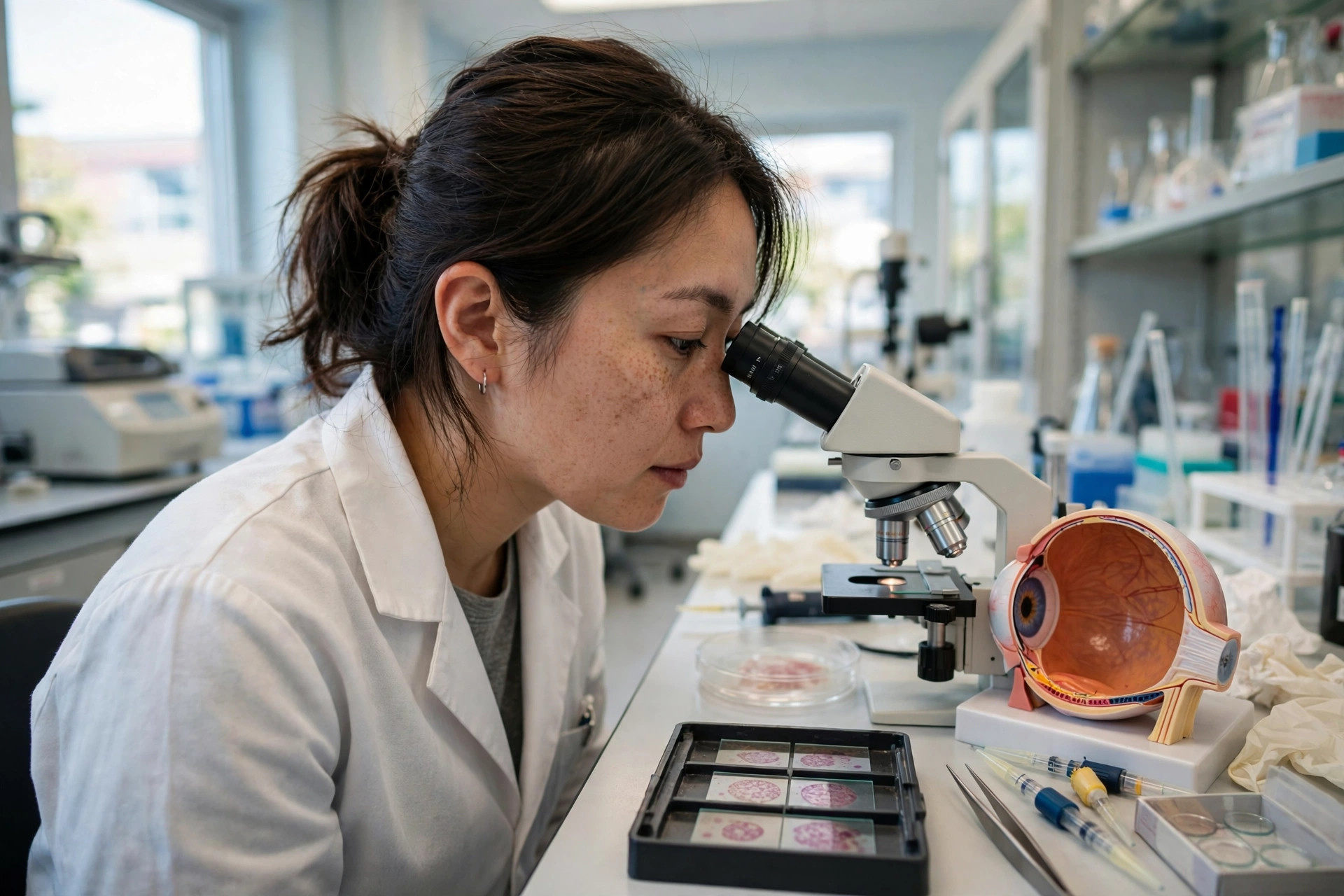
A hypoxia-signal stabiliser protected light-sensing cells and raised mitophagy markers — in mouse cells and retinal detachment
When the retina detaches, photoreceptor cells lose their oxygen supply and begin to die — the race in retinal detachment surgery is against that cell death. A study in Cell Death & Disease (published November 7, 2026) tested a new way to switch on the cell's own hypoxia defence: Stabilizer 1a, a cyclometalated iridium(III) complex that disrupts the VHL-HIF1alpha interaction, stabilizing HIF1alpha — the master transcription factor of oxygen adaptation — even when oxygen is present. In the murine cone photoreceptor-like 661W cell line, 1a increased HIF1alpha under both normal and low oxygen, outperformed the comparison agent DMOG on the reported readouts, reduced hypoxia-induced apoptosis (less cleaved caspase-3 and annexin V), reduced mitochondrial and total reactive oxygen species, preserved ATP production, and increased mitophagy markers — while also inducing a reversible senescence-like state (raised p16 and p21, reduced proliferation; removing the compound restored p21 to baseline). In living mice, 1a attenuated photoreceptor apoptosis after experimental retinal detachment, again with increased mitophagy markers. The abstract reports these effects qualitatively, without percentage sizes.
Read article →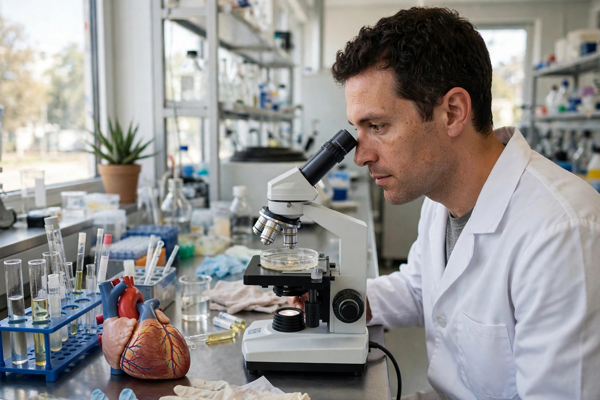
Silencing one gene protected heart cells from high-glucose mitochondrial injury — in human cardiomyocytes
Diabetic heart muscle fails in part at the mitochondria: quality control — the import, repair and mitophagy systems that keep the organelle pool healthy — breaks down under chronic high glucose. A study in Frontiers in Cardiovascular Medicine (published November 9, 2026) mined a rat diabetic-myocardium dataset (GSE4745) plus three supportive human and animal cohorts and identified 192 differentially expressed genes, 58 of them tied to mitochondrial quality control, with altered fatty-acid oxidation, mitochondrial protein import and mitophagy programs. One candidate stood out: proenkephalin (PENK), better known as a precursor of opioid peptides. PENK rose under high glucose, and in human AC16 cardiomyocytes treated with high glucose, silencing PENK with either of two independent siRNAs improved cell viability, reduced cleaved caspase-3 and mitochondrial superoxide, restored the JC-1 membrane-potential ratio, lowered PINK1, Parkin and DRP1, and raised the fusion protein MFN2. The abstract reports these effects qualitatively, without percentage effect sizes — a reason to read this as a promising lead, not a measured therapy.
Read article →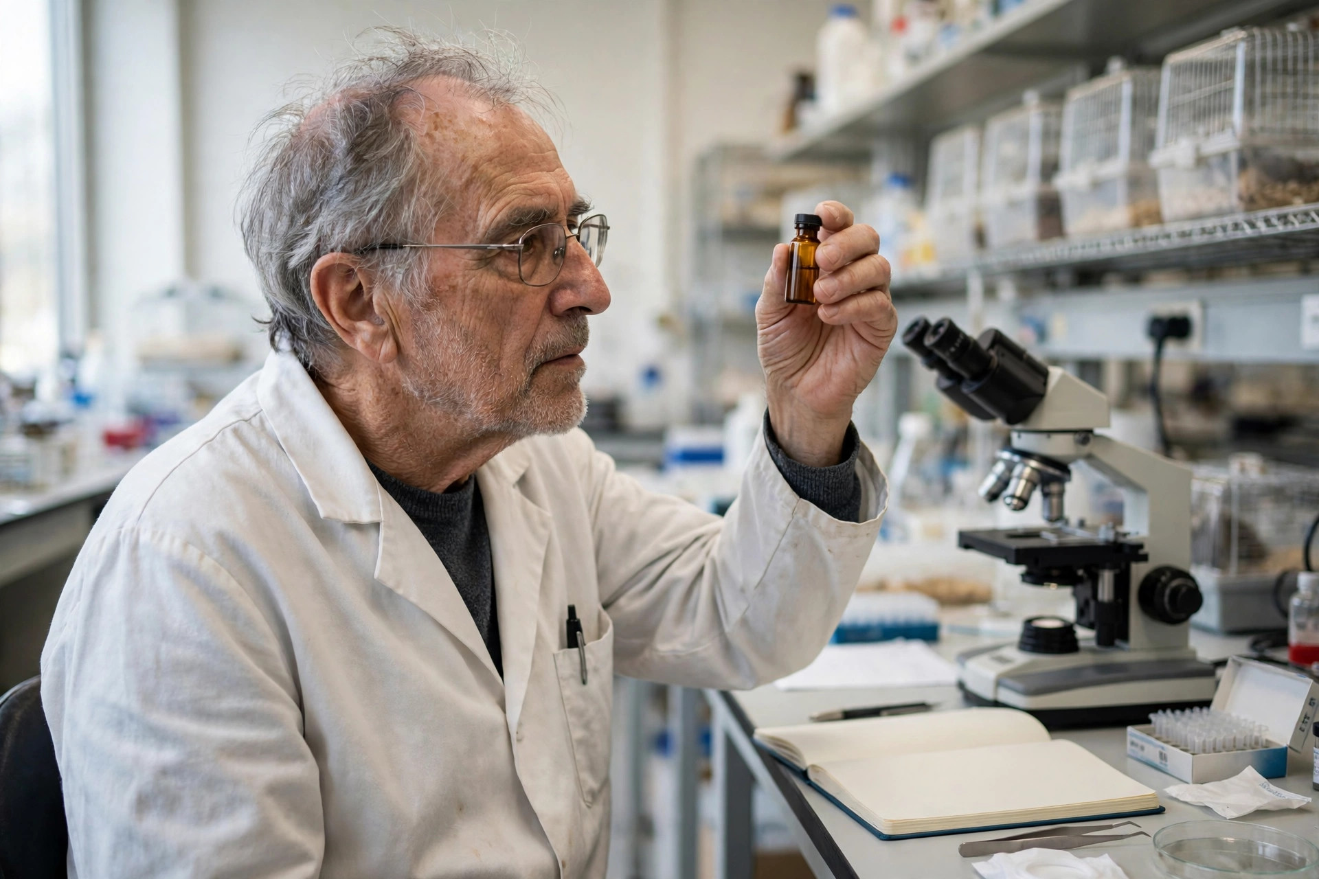
A blood-sugar mineral cleared senescent cells and extended lifespan — in mice
Senescent cells — aged cells that stop dividing but refuse to die — accumulate with age and drive tissue decline, and selectively removing them (senolysis) is a central longevity strategy. A study in Nature Aging (Wu, Yang, Li and colleagues, University of Chicago and collaborators; published November 6, 2026) found that senescent cells carry an altered pattern of reactive protein cysteines, and that this chemistry is a vulnerability: vanadyl sulfate, a vanadium compound long sold as a blood-sugar supplement, selectively oxidizes those cysteines, driving catastrophic protein aggregation and autophagic death in senescent cells while sparing healthy cells. In aged male mice given vanadyl sulfate in drinking water for three months starting at about 17 months of age, median survival rose to 952 days versus 886.5 days in controls — 65.5 days longer — alongside doubled hanging grip endurance, less visceral fat, and reversed hepatic and renal decline markers in the phenotype experiments. The caveats are substantial and stated plainly below: mice only, a males-only lifespan arm, a narrow therapeutic window for vanadium, and a mouse dose far above typical supplement doses. This is not a recommendation to take vanadyl sulfate.
Read article →
Driver or amplifier? A new review grades where mitochondria truly cause brain disease — and where they pile on
Mitochondrial dysfunction appears in nearly every neurodegenerative disease — but appearing is not causing. A 60-page review published November 7, 2026 (IMU University and colleagues, International Journal of Molecular Sciences) set out to grade the evidence disease by disease for Alzheimer's (AD), Parkinson's (PD), ALS and Huntington's (HD), weighing human-genetic, patient-tissue and model-based evidence separately. Its verdict: mitochondrial dysfunction is best supported as a primary driver in Parkinson's disease and in genetic ALS, and as an early amplifier of protein-driven or mutant-huntingtin toxicity in sporadic Alzheimer's and Huntington's. The review walks the shared machinery — defective oxidative phosphorylation, altered fission–fusion dynamics, defective mitophagy, impaired axonal transport, excess reactive oxygen species, and the cell-death pathways they feed (necroptosis, ferroptosis, pyroptosis, parthanatos) — and the disease-specific signatures: amyloid-beta/tau impairing mitophagy in AD, PINK1/Parkin dysfunction and complex I inhibition in PD, SOD1/C9orf72-associated defects in ALS, mutant huntingtin's bioenergetic effects in HD. It is equally direct about translation: despite the mechanistic richness, mitochondria-targeted therapies have not yet succeeded clinically — while noting newer approaches including USP30 inhibition, intercellular mitochondrial transfer, and the regulatory approval of a mitochondria-targeted peptide.
Read article →
Brain tissue grown from PSP patients lost its energy reserve — a coordinated mitochondrial defect, in lab-grown human organoids
Progressive supranuclear palsy (PSP) is a primary 4-repeat tauopathy: deposits of an abnormal tau protein accumulate in vulnerable neurons, and the Richardson's syndrome form (PSP-RS) is its most common and devastating presentation. A study in Cellular and Molecular Life Sciences (Lazzinnaro, Esposito, Parrotta and colleagues; published November 3, 2026, with fresh coverage November 8) grew midbrain organoids — laboratory-grown human brain tissue — from PSP-RS patients and mapped how tau pathology connects to mitochondrial dysfunction, combining functional bioenergetic analysis, quantitative proteomics, targeted metabolomics and redox imaging. The central finding concerns respiratory reserve: PSP-RS organoids showed a marked reduction in maximal respiration and spare respiratory capacity, the buffer cells draw on when energy demand surges. The authors describe a coordinated phenotype — respiratory chain remodelling alongside oxidative stress and metabolic reprogramming — and are explicit about scope: mitochondrial dysfunction appears as an integrated feature of PSP-RS in this model; the work does not identify a cause of the disease or a therapeutic target, and organoids are a model system, not measurements in living patients' brains.
Read article →
A ginseng compound extended lifespan and protected neurons — in worms, by restoring the cell's recycling system
Lysosomes are the cell's recycling centres: they digest worn-out proteins and damaged organelles — including worn-out mitochondria, through mitophagy — and their decline is a hallmark of aging. A study in Aging Cell (Yu, Li, Chen and colleagues; published November 7, 2026) tested 20(S)-ginsenoside Rg3, a compound from ginseng, in the roundworm C. elegans. At 10 micromolar, Rg3 extended lifespan and preserved the functions that normally fade with age — locomotion and pharyngeal pumping — and it worked when given only in adulthood, though the benefit was strongest when treatment began early in life. It also maintained mechanosensory neuron integrity and touch sensitivity, suppressed neuronal polyglutamine aggregation, and protected dopaminergic neurons against alpha-synuclein degeneration. Mechanistically, Rg3 restored neuronal lysosomal function and autophagic flux: it shifted phosphoinositide lipids at lysosomal membranes (PI(4)P up, PI(3)P down), reduced RSKS-1/S6K phosphorylation — a sign of suppressed TORC1 signalling — and drove HLH-30, the worm version of the lysosomal master switch TFEB, into the nucleus. The causal controls are unusually complete: knocking down bec-1 (a core autophagy gene), vha-3 (the lysosomal proton pump) or hlh-30 abolished the benefits. Every result is in worms; there are no human data.
Read article →
When muscle inflames, its mitochondrial DNA may be both casualty and culprit — a new evidence review
The idiopathic inflammatory myopathies (IIMs) — immune-driven muscle diseases including dermatomyositis, polymyositis and inclusion body myositis — pair muscle weakness with inflammation, and mitochondria sit at the crossing point. A narrative review published November 6, 2026 (Second Hospital of Hebei Medical University and colleagues, International Journal of Molecular Sciences) integrates what is known about mitochondrial DNA abnormalities in these diseases. Large-scale mtDNA deletions and depletion are documented in inclusion body myositis and in polymyositis with mitochondrial pathology, while dermatomyositis studies show regional depletion and oxidative modifications in distinct tissue compartments. The mechanistic thread: mtDNA encodes essential oxidative-phosphorylation components, and when it escapes damaged mitochondria it can stimulate innate immune sensing and inflammasome activation — potentially feeding the very inflammation that injures the muscle. The review is candid about the therapeutic gap: reported clinical benefits of mitochondria-directed approaches do not establish mtDNA-specific therapeutic effects, and it names subtype-by-subtype biomarker validation as the field's priority. It is a synthesis of prior work (searches through October 20, 2026), not a new experiment.
Read article →
A traditional plant extract protected aging hearts' mitochondria — in mice and lab-grown heart cells
Smilax glabra Roxb. is a rhizome used in traditional medicine, and its flavonoid fraction (SGF) had already shown protection against pathological cardiac hypertrophy. A study published November 6, 2026 (Zhejiang Academy of Traditional Chinese Medicine and colleagues) asked whether SGF protects against aging-like cardiac change, using D-galactose — a sugar that induces aging-like damage — in C57BL/6J mice and in H9c2 heart cells, with mitochondrial structure as a central readout. In the mice, SGF significantly attenuated cardiac hypertrophy, reduced myocardial fibrosis and reactive-oxygen-species overproduction, and preserved mitochondrial ultrastructure, alongside increased AMPK phosphorylation and higher expression of the mitochondrial-biogenesis axis PGC-1alpha/NRF1/TFAM and the mitophagy pathway PINK1/Parkin. In the cells, SGF suppressed senescence-like changes — and those benefits were abolished when an AMPK inhibitor (Compound C) was added, with SGF's action resembling the AMPK activator AICAR. The authors frame the mechanism carefully: an AMPK-associated pattern that may contribute to the cardioprotection, in an artificial aging model, with no human data.
Read article →
Long-chain PFAS replacement chemicals impaired placental cells' mitochondria — in lab-grown human trophoblast cells
When regulators squeezed the best-known 'forever chemicals,' industry shifted to alternatives — including hexafluoropropylene oxide (HFPO) homologues, now increasingly detected in environmental and biological samples. A study in Environment & Health (Wei and colleagues; published November 6, 2026) tested whether chain length decides toxicity in HTR-8/SVneo cells, a human trophoblast-like line used to model the placenta's invasive cells. The long-chain compounds HFPO-TA and HFPO-TeA — but not short-chain HFPO-DA or PFOA — suppressed proliferation, arrested the cell cycle at S phase, and impaired migration and invasion. Transcriptomic profiling pointed to disrupted integrin-mediated adhesion and focal-adhesion signalling, with FAK/PI3K/Akt signalling inhibited, alongside mitochondrial dysfunction and excess reactive oxygen species. The mitochondrial numbers are concrete: membrane potential fell by 19% and 31% under 50 and 100 micromoles per litre of HFPO-TA, and by 12%, 24% and 73% under 25, 50 and 100 micromoles per litre of HFPO-TeA, while intracellular ROS rose up to 1.8-fold and 2.7-fold respectively. Molecular docking suggested preferential interactions between HFPO-TeA and integrin subunits. Every result is in cultured cells at laboratory concentrations.
Read article →
PD-L1, the immunotherapy target, tracks with mitochondrial remodelling in lung cancer cells — in lab-grown cells
PD-L1 is famous as the brake pedal cancers press to evade the immune system — and the target of blockbuster immunotherapies. A study in Cell Death & Disease (Shi, Liu, Mou and colleagues, Beijing Tiantan Hospital and Capital Medical University with Tsinghua University; published November 4, 2026, as an accepted early version) asks what else PD-L1 status tracks with inside the cancer cell itself. Comparing non-small cell lung cancer (NSCLC) cells by PD-L1 expression, the team found PD-L1 associated with increased endocytic activity and altered metabolic flux, particularly in the tricarboxylic acid (TCA) cycle — the mitochondrial energy hub. PD-L1 expression was also associated with mitochondrial remodelling: changes in proteins governing mitochondrial fusion and fission and in respiratory-chain components. Integrated multi-omics analyses further found differential H3K9me3 epigenetic patterns by PD-L1 status, including at the PTPRN2 locus. Every finding is an association in cultured cells; no treatment was tested and no direction of cause is established.
Read article →
A lactate-driven tag on a mitochondrial enzyme protected blood vessels in diabetic plaque models — in mice and endothelial cells
IDH2 is a mitochondrial enzyme that helps make alpha-ketoglutarate (alpha-KG), a central metabolic intermediate. A study in Cell Death & Disease (Xu, Zang, Wang and colleagues, Jiangsu University, corresponding author Zhongqun Wang; published November 5, 2026) reports that a chemical tag called lactylation — placed on IDH2 at one specific site, lysine 272 — is significantly elevated in diabetic arterial plaques and acts as a protection switch. Lactylation at K272 maintained IDH2's enzymatic activity, keeping alpha-KG production sufficient and attenuating mitochondrial damage and endothelial dysfunction under high-glucose, high-lipid conditions. In two different mouse models engineered so the tag could not be placed, losing K272 lactylation worsened mitochondrial dysfunction and endothelial injury and promoted plaque formation and instability. Supplementing alpha-KG itself effectively mitigated diabetic plaque progression in the model. The abstract reports no effect sizes.
Read article →
Restoring ITGA5 cleared damaged mitochondria and sped diabetic wound healing — in diabetic mice, human wound tissue and lab-grown endothelial cells
Diabetic foot ulcers stall, in part, because the blood-vessel cells that should rebuild tissue are choking on damaged, iron-loaded mitochondria. A study in the Journal of Translational Medicine (Sun, Yuan, Zhang, He and Xiong, Xiang Xiong's group at the Second Xiangya Hospital, Central South University; published November 4, 2026) found the adhesion protein ITGA5 (integrin subunit alpha 5) significantly reduced in human diabetic foot ulcer tissue compared with normal skin. Restoring ITGA5 in db/db diabetic mice — via lentiviral overexpression — accelerated wound closure, improved collagen organization and increased new blood-vessel formation. In endothelial cells stressed by high glucose, ITGA5 restoration improved survival, migration and tube formation, while removing ITGA5 made things worse: more mitochondrial reactive oxygen species and more mitochondrial iron overload. Mechanistically, ITGA5 switched on PI3K/AKT signaling, cut mitochondrial iron accumulation and lipid peroxidation, and switched on a disposal route called mitocytosis — damaged mitochondria packaged into migrasomes and expelled from the cell.
Read article →
A helicase that supercharges mitochondrial energy production drives colorectal cancer growth — in human tumour tissue, cancer cells and mice
Colorectal cancers lean hard on mitochondrial oxidative phosphorylation (OXPHOS) — the cell's main energy assembly line. A study in Cell Death Discovery (He, Zhang, Xu and colleagues, Ji Tao's group at Harbin Medical University Cancer Hospital; published November 3, 2026) identifies the DEAD-box helicase DDX49 as a previously unrecognized regulator of that dependence. DDX49 was abnormally upregulated in colorectal cancer tissues, and higher expression tracked with more advanced stage and worse prognosis. In the laboratory, DDX49 promoted colorectal cancer cell proliferation and tumour growth and suppressed apoptosis. The mechanism runs straight through the mitochondria: nuclear DDX49 binds and stabilizes PGC-1α — the master switch of mitochondrial biogenesis — which raises TFAM and OXPHOS complex subunits and increases mitochondrial respiration. Rescue experiments confirmed PGC-1α is an essential downstream mediator. The authors propose DDX49 as a prognostic biomarker and a therapeutic target; no drug against it was tested.
Read article →
Smoking-linked cadmium switches on a brain signal that damages neuronal mitochondria — in human data and lab-grown neurons
Cadmium is a toxic heavy metal with no job in the body, and smoking is one of its main delivery routes. A study published November 3, 2026 in the Journal of Translational Medicine (Peng, Zhou, Guo and colleagues, Guizhou Medical University) traced a chain from that exposure into the Alzheimer's brain. Integrating survey-weighted NHANES population data, the team confirmed that ever-smoking was associated with higher whole-blood cadmium concentrations. Layering transcriptomic and single-nucleus analyses on top nominated a candidate mechanism: MAPK9, the gene for the stress kinase JNK2, enriched in excitatory neurons in connection with amyloid-beta-linked mitochondrial complex I dysfunction. In human brain tissue, JNK2 phosphorylation was increased in Alzheimer's disease. In neurons, cadmium chloride raised JNK2 phosphorylation and the abundance of APP — the amyloid precursor protein — and impaired mitochondria. Molecular docking supported the plant compound sanguinarine binding MAPK9 stably and mitigating the mitochondrial dysfunction in their models. This is an association-and-mechanism study: it nominates a pathway (the smoking–cadmium–MAPK9 axis) rather than proving cadmium causes Alzheimer's, and sanguinarine is a docking-supported candidate, not a treatment.
Read article →
In newborn brain injury, one cell-death pathway splits in two: one arm summons mitochondrial cleanup, the other causes the damage — in mouse neurons and a neonatal mouse model
When a newborn's brain is starved of oxygen — hypoxic-ischaemic encephalopathy (HIE), a devastating injury — two cleanup-and-death systems switch on together, and a study published November 3, 2026 in Cell Death & Disease (Wu, Chang, Zhao and colleagues, Daqing Ma's group at Imperial College London and collaborating institutions in China) shows they are not one system but a fork. Using oxygen-glucose deprivation and reperfusion in mouse primary neurons and Neuro-2a cells, the team found the injury activated necroptosis — a programmed cell-death pathway — with necrosome components moving onto mitochondria, alongside induction of mitophagy, the mitochondrial quality-control cleanup (Parkin and BNIP3 upregulated, mitochondrial LC3 conversion increased, the mitochondrial protease ClpP induced). Blocking RIPK1 or RIPK3, the pathway's upper kinases, attenuated the mitophagy — those arms promote the cleanup response. But knocking down MLKL, the pathway's executioner protein, did the opposite: it enhanced mitophagy and restored mitochondrial function and morphology. A neonatal mouse model of HIE showed both pathways activating together in the hippocampus. The practical upshot is a targeting warning: drugs that block this pathway wholesale could suppress a protective cleanup along with the damage.
Read article →
Gut bacteria make fats that feed liver and muscle mitochondria — in mice
Some of the signals that tune your mitochondria are not made by you at all. An interdisciplinary team from UConn and the University of Pittsburgh reports two linked studies (Gastro Hep Advances and The Journal of Physiology, 2026; reported November 3, 2026) on glycine lipids — fats produced by Bacteroidota species that live in the gut and mouth. The lipids circulate through the body: the team detected them in both portal blood and intestinal lymph, and in human liver tissue samples. In mice fed a high-fat, high-sugar Western diet that drives metabolic-dysfunction steatotic liver disease (MASLD), one-microgram injections of glycine lipids L654 or L1256 every other day for eight weeks significantly attenuated liver disease severity, switched on genes controlling mitochondrial biogenesis and fat oxidation, and depleting the lipids worsened inflammation while restoring them brought it back to healthy levels; a high-fiber diet (pea fiber plus citrus peel fiber) raised the lipids' levels. In a companion study in male mice on a Western diet, L1256 accumulated in skeletal muscle and improved fatty-acid-supported mitochondrial respiration, increased proteins for energy production and antioxidant defense, and helped preserve muscle fiber size. All efficacy data are in mice and cells — the human data so far show only that the lipids are present in human liver — and a human fiber-intervention study is being started, not reported.
Read article →
Mitochondrial disease makes the body burn more fuel to do less — in a small human study
It sounds backwards: people whose cells struggle to make energy turn out to spend more of it. A study in Cell Press Blue (Shaulson, Sercel, Rausser and colleagues, Columbia University with the CU Anschutz Health and Wellness Center; reported November 3, 2026) compared 10 adults with mitochondrial disease to 10 healthy adults and found context-specific hypermetabolism: under controlled laboratory conditions, the mitochondrial-disease group expended approximately 195 additional calories per day, even while moving less — with elevated energy expenditure particularly during sleep. In everyday life the picture inverted: total daily energy expenditure, measured with the doubly labeled water gold-standard method, was similar between groups, because the mitochondrial-disease group was approximately 39 percent less physically active, as if reduced activity compensates for higher underlying energy costs. The mitochondrial stress protein GDF15 was nearly five times higher in the mitochondrial-disease group and was associated with greater fatigue, higher energy demands and reduced activity. Ten people per group is a small study, and hypermetabolism was context-specific rather than constant — but this is direct human measurement of a question usually argued from cell biology.
Read article →
Switching on the cell's own protein shredders restored mitochondrial function — in lab-grown human cells
Most drug hunters destroy one bad protein at a time. A study published in Science (Chandrasekhar, McMinimy, Manford and colleagues, Michael Rapé's group at UC Berkeley; online October 29, 2026) demonstrates the opposite move — stimulated protein degradation (SPD): instead of dragging one target to the cell's shredder, a small molecule takes the brakes off the shredder itself. The team targeted CUL2FEM1B, an E3 ubiquitin ligase that guards mitochondria by degrading proteins that block mitochondrial protein import, including FNIP1. Under normal conditions, BEX-family proteins sit in the ligase's substrate pocket and restrain it. Screening roughly 120,000 compounds produced BD1, then a cell-active analogue, BD2, which displaces the BEX brakes and lets CUL2FEM1B degrade several of its natural substrates at once. In cultured-cell models of cytochrome c oxidase (COX) deficiency — where loss of assembly factors had cut respiratory capacity by 30 to 40 percent — BD2 restored respiration and prevented the death of neurons grown from induced pluripotent stem cells (iPSCs), and in frataxin-depleted models of Friedreich's ataxia it matched the effect of the approved drug omaveloxolone. It did not rescue every defect tested, and every result is in cultured cells — no animals, no patients, no pharmacology yet.
Read article →
A £2.1 million bet on brain mitochondria: Cardiff wins a Wellcome award to hunt the genes that run them, in fruit flies
Dementia's energy problem just got funded like a first-order question. Cardiff University announced (October 28, 2026; shared widely November 2) that Dr Gaynor Smith, a Reader at the UK Dementia Research Institute at Cardiff, has received a £2.1 million Wellcome Discovery Award for a project using fruit flies to study how mitochondria — the cell's energy-producing structures — are genetically regulated in brain cells. The premise, in Smith's words: neurons require a constant supply of energy, largely generated by mitochondria, which are highly dynamic structures that continually move through cells, change shape, divide and fuse to meet the demands of healthy brain function; mitochondrial dysfunction is increasingly recognised as a key feature of several neurodegenerative diseases, yet many of the genes and pathways controlling mitochondrial behaviour remain poorly understood. The project will investigate how brain cells stay healthy and what happens when the systems that power them stop working properly, aiming at insights into Alzheimer's and Parkinson's disease. This is a funding announcement, not a result: no findings are reported, and fruit-fly genetics is a discovery engine whose hits still have to survive translation into mammalian and human biology.
Read article →
Aging rewrites the fat in every tissue differently — but one mitochondrial lipid moves everywhere, in rats
If mitochondria are engines, their membranes are built of fat — and aging rebuilds the fat. A study published November 2, 2026 in Scientific Reports (Aisin, Sokolova, Grishina and colleagues) ran untargeted LC-MS lipidomics on five compartments from young and old male Wistar rats — frontal cortex, cerebellum, liver, muscle and plasma — quantifying 884 lipids across 36 classes. Muscle changed the most, with enrichment of PG and PE O- lipids indicating mitochondrial dysfunction. Plasma shifted in phosphocholine lipids (LPC and PC O-); the liver unexpectedly lost triglycerides, particularly shorter- and odd-chain species, consistent with reduced gut-microbiota-derived fatty acids; brain regions showed myelin-related changes. No single lipid changed in every tissue — aging's lipid signature is local. But at the class level one signal coordinated across tissues: PG (phosphatidylglycerol, the precursor family of the mitochondrial signature lipid cardiolipin) showed the strongest coordinated age-related changes, which the authors read as mitochondrial lipid remodeling being a common aging feature. These are rats, descriptively profiled — no intervention, no human samples, and association between lipid patterns and dysfunction rather than a test of cause.
Read article →
Neurons need their mitochondria to prune their own branches — in fruit flies
Growing brains sculpt themselves by cutting: neurons prune excess branches so adult circuits wire correctly. A study published today, November 3, 2026, in Scientific Reports (Mukherjee, Cheng, Sproston and colleagues, Darren Williams' group at King's College London) watched that cutting happen in living fruit-fly sensory neurons and found mitochondria on the required-crew list. Using a sensitive dual-colour probe (SR4VH), the team saw executioner caspase activity — the molecular scissors of cell death, used here at sub-lethal levels — switch on in the pruning neuron ddaC within 20 minutes of puparium formation, well before its larval dendrites were severed, and at consistently lower levels than in neighboring neurons that were actually dying. Single-cell mutant clones showed the pro-death RHG proteins are required inside the pruning cell itself, with reaper and grim — rather than hid — cooperating to drive branch severing. And when mitochondrial function or transport was disrupted, dendritic mitochondria were depleted, caspase activation was prevented, and both pruning in ddaC and apoptosis in its neighbors were blocked. This is basic developmental biology in flies — it tests no therapy and studies no human brain — but it pins down, in an intact animal, how neurons borrow death machinery without dying.
Read article →
When a DNA-repair pathway fails, developing brain cells lose their mitochondria too — in lab-grown human cells
Fanconi anemia is known as a bone-marrow failure disorder, but its brain effects have never had a clear mechanism. A study published November 2, 2026 in Cell Death & Disease (Shi, Dong, Wu and colleagues, Lei Li's group at Zhejiang University) shows that in human cellular models, losing the Fanconi anemia pathway selectively derailed neuron and glia development: neuronal shape was impaired, neuron and oligodendrocyte markers were defective, mitochondria malfunctioned, and the cells' electrical activity was faulty. The trigger the team traced is strikingly ordinary: as cells reprogrammed their genes to become neurons, a metabolic surge of endogenous formaldehyde — a chemical the body itself makes — built up DNA-protein crosslinks and drove cell death in cells lacking the FANCL gene. Restoring FANCL, or restoring ADH5, the enzyme that detoxifies formaldehyde, rescued the defects. Media from the affected cultures also provoked a reactive, inflammation-like response in healthy recipient cells, suggesting damage spreads beyond the cells that carry the defect. All of this is in lab-grown human cell models — no patients were treated, and no therapy was tested.
Read article →
A mitochondrial enzyme guards bone-building cells as they age — losing it sped bone loss in mice
Bone loss with age is usually told as a calcium story. A study published October 31, 2026 in Cell Death Discovery (Song, Huang, Guo and colleagues) tells it as a mitochondrial quality-control story. The team mapped a three-protein axis — PRMT6, SIRT5 and HADHA — in ageing bone. SIRT5 is a mitochondrial desuccinylase: it removes succinyl chemical tags from proteins, and one of its targets is HADHA, a core subunit of the mitochondrial machinery that burns fatty acids. In mice lacking Sirt5, succinyl tags accumulated globally, HADHA was tagged at multiple sites and lost, and osteoblasts — the cells that build bone — accumulated R-loops, tangled DNA-RNA structures around the start sites of genes, along with DNA damage. Compared with normal mice, Sirt5-deficient mice showed more pronounced trabecular bone loss and impaired bone formation, with reduced Runx2, ALPL and OCN (bone-building markers) and elevated p16 and p21 (senescence markers). Losing Prmt6 lowered Sirt5 and copied the same pattern. The rescue ran the other way: delivering extra Sirt5 with an AAV5 viral vector restored HADHA, reduced R-loops and senescence, and alleviated trabecular bone loss. Everything causal here is in mice and cultured bone cells — there is no human bone data in the paper.
Read article →
In kidney disease, phosphate flips a glutamine switch that strains heart mitochondria — and blocking it protected hearts in mice
Heart enlargement is one of the deadliest complications of chronic kidney disease, and this study traces part of it to a metabolic switch inside heart-muscle mitochondria. In a paper published November 1, 2026 in Nature Communications, Bai, Wang, Zhou and colleagues (Junnan Wu's group at Zhejiang University and Sir Run Run Shaw Hospital) integrated transcriptomic, metabolomic and proteomic analyses and identified phosphate — which builds up in kidney disease — as a major regulator of glutaminase 1 (GLS1), the mitochondrial enzyme that controls glutamine burning. Excessive GLS1 activation increased ammonia production, disrupted mitochondrial respiratory protein expression and redox homeostasis, and reduced bioenergetic efficiency. In male mice with kidney disease, deleting GLS1 only in heart-muscle cells, or inhibiting it with a drug, attenuated left ventricular hypertrophy. Human data point the same way but prove less: in a multicenter peritoneal dialysis cohort, serum-induced GLS1 activity was associated with hypertrophic remodeling, and human heart samples from people with kidney disease showed increased GLS1 expression. The intervention evidence is mouse-only, in males; the human evidence is association, not treatment.
Read article →
Moving cancer cells shed giant vesicles carrying whole genomes and working mitochondria — in lab-grown cells
Cancer cells on the move leave something behind that cell biology had not catalogued. A study published October 30, 2026 in Nature Cell Biology (Tsering, Burnier, Lyden and colleagues, McGill University and Weill Cornell Medicine) describes 'motile vesicles' — MoVes — large vesicles, 2 to 5 micrometres across, budded from the trailing edge of migrating cancer cells and left anchored on the surface behind them. Pooled MoVes material carried the parent cell's entire genome and mutational landscape, and the vesicles contained organelles including mitochondria, Golgi and endoplasmic reticulum. MoVes appeared in highly migratory breast cancer lines, were absent from non-cancerous breast epithelial cells until an oncogene made them mobile, and their production rose sharply when the invasive program called epithelial-to-mesenchymal transition was switched on. Their mitochondria kept an intact membrane potential, and in dish experiments MoVes raised respiration and mitochondria-dependent ATP production in recipient cells stripped of their own mitochondrial DNA. This is a laboratory characterisation of a new vesicle type — not a test for cancer spread and not evidence of what MoVes do inside a patient.
Read article →
In a genetic heart disease, mitochondria change before symptoms do — and a plant compound blunted the shift, in mice
Arrhythmogenic cardiomyopathy (ACM) is an inherited heart-muscle disease, most often caused by mutations in PKP2, the gene for the desmosomal protein plakophilin-2, and its most dangerous phase is the early 'concealed' stage, when fatal rhythm disturbances can strike before any symptom. A study published October 29, 2026 in Nature Cardiovascular Research (Achter, Svenningsen, Mulvey, Bertoli and colleagues, in the Lundby and Delmar laboratories) used time-resolved proteomics in mice whose heart cells could be switched to lose Pkp2 in adulthood: across 72 heart samples and more than 6,500 proteins measured at five time points over 21 days, mitochondrial remodeling was among the earliest detectable changes — mitochondria expanded while key metabolic machinery was selectively lost, with oxidative stress arriving later. Right-ventricle mitochondria, unlike left, could not raise ATP production in response to calcium. Treating the mice for 14 days with honokiol, a magnolia-bark polyphenol reported to engage the mitochondrial enzyme sirtuin-3, shifted protein abundances broadly back against the disease trajectory. All of this is in a mouse model of the disease's first three weeks — a map of where to intervene, not a treatment result.
Read article →
In sepsis, the injured kidney ships mitochondrial DNA to the lungs — and the lungs are injured by the delivery, in mice and human data
Sepsis kills by spreading: one organ fails, then another, and how the damage travels has been a standing puzzle. A study published October 30, 2026 in Communications Biology (Jiao, Zhang, Xie and colleagues, Nanjing) identifies a courier. The injured kidney packages mitochondria-rich extracellular vesicles — mitoEVs — into the blood; the vesicles travel to the lung, are taken up by its blood-vessel lining cells, and the mitochondrial DNA they carry switches on the cGAS-STING innate immune pathway, disrupting the endothelial barrier, recruiting immune cells and driving respiratory failure. In septic mice, sepsis switched on vesicle formation in kidney tubule cells by raising the protein sorting nexin 9, and blocking that process in the kidney reduced mitoEV release and protected the lungs. In patients with sepsis, higher circulating mitoEV levels were associated with lung injury and death. The mechanistic chain is from mice and cultured cells; the human data are an association, not proof of the same pathway in people.
Read article →
A four-protein assembly line builds two of the cell's energy machines at once — in yeast, with a human counterpart
Mitochondria do not import their biggest machines ready-made; they assemble them on site, and the assembly crews are still being discovered. A paper published in EMBO Reports (Pedroza-Dávila, Perez-Martinez and colleagues) characterizes a four-protein complex in baker's yeast, named AMIGa, that coordinates construction of both cytochrome c oxidase — complex IV, the final step of the respiratory chain — and ATP synthase, the enzyme that makes most of the cell's ATP. The inner-membrane protein Aim11 works with partners Mtc3, Gep7 and Iai11 at the crossroads of the two assembly lines. Basic science earns a place in this edition for one specific reason: the team identified the human protein TMEM242 as Aim11's counterpart, and TMEM242 compensated for the loss of Aim11 in yeast — a direct thread from a yeast assembly crew to human mitochondrial biology, where assembly defects cause real disease.
Read article →
A mitochondrial enzyme drives chemotherapy heart damage by leaking damaged DNA — and a vitamin coenzyme blocked it, in mice
Doxorubicin saves lives in cancer care, but its dose-limiting harm is damage to the heart. A mouse study published October 29, 2026 in Cellular & Molecular Biology Letters (Feng, Xie and colleagues) identifies the mitochondrial enzyme CMPK2 as a central driver: switched on by the stress transcription factor STAT1, CMPK2 drives oxidative damage to mitochondrial DNA, that damaged DNA leaks into the cell body, and the leak triggers the NLRP3 inflammasome — the inflammatory complex that injures heart muscle. In cardiomyocyte-specific knockout and overexpression mice, removing CMPK2 protected the heart and forcing it worsened injury. Most strikingly, flavin adenine dinucleotide (FAD) — the active coenzyme form of riboflavin, vitamin B2 — blocked the damaging pathway in mice without reducing doxorubicin's antitumor effect. Every result here is preclinical: mice and cell assays, no human data.
Read article →
An RNA-modifying enzyme drives egg-follicle loss by blocking mitophagy — and blocking it protected follicles, in animal and cell studies
Most eggs a woman is born with are lost long before ovulation, in a process called follicular atresia — and the support cells around the egg, granulosa cells, decide much of that fate. A paper published October 25, 2026 in Cell Death Discovery (Zhou and colleagues) traces a chain from an RNA chemical modification to mitochondrial failure: the enzyme NAT10 adds an ac4C modification to the messenger RNA for the protein Tnc, stabilizing it; the downstream effect suppresses Parkin-mediated mitophagy, the pathway that clears damaged mitochondria, so oxidative stress builds in granulosa cells and follicles die. Interrupting the chain worked in the models tested: the NAT10 inhibitor Remodelin promoted mitophagy and reduced oxidative stress in granulosa cells in vitro, and in vivo it hindered follicular atresia and improved reproductive efficiency impaired by granulosa-cell oxidative stress. The authors present NAT10 as a therapeutic candidate target for infertility and subfertility. The study is an accepted, peer-reviewed manuscript published ahead of its final version of record — preclinical throughout, with no human data.
Read article →
The heart imports lactate straight into its mitochondria to keep beating under stress — in mice and human heart cells
For decades, the textbook route for carbohydrate fuel was simple: glucose becomes pyruvate in the cell body, and pyruvate enters mitochondria through the mitochondrial pyruvate carrier. A paper published October 28, 2026 in Nature (Cluntun, Visker, Ducker and colleagues, University of Utah) reports a parallel route that bypasses that gate. In mouse heart muscle, glucose was still oxidized even when the pyruvate carrier was genetically removed — because the transporter MCT1, found in the inner mitochondrial membrane of both mouse and human heart cells, imports lactate directly into mitochondria, where the enzyme LDHB converts it to pyruvate and NADH. When MCT1 was removed instead, mitochondrial lactate oxidation stopped, the heart's adaptation to stress failed, and heart failure accelerated. Senior author Gregory Ducker's summary: this appears to be a critical backup system for heart function — mostly dispensable in daily life, crucial when the heart is stressed.
Read article →
Two mitochondrial proteins moonlight as disposal tags for damaged mitochondria — and their failure tracks a nerve disease, in cell studies
Mitofusin 2 (MFN2) is famous for fusing mitochondria together, and SLC25A46 for shaping them. A PNAS paper published October 27, 2026 (University of Zurich researchers) gives both proteins a second job: when a mitochondrion is damaged, MFN2 and SLC25A46 bind the autophagy machinery proteins LC3 and GABARAP directly on the organelle's surface, acting as mitophagy receptors that flag it for disposal. In fibroblasts from Charcot-Marie-Tooth disease patients carrying MFN2 mutations, that clearance failed — damaged mitochondria accumulated and neuronal survival under stress fell. The work is in cells, not patients, but it reframes a disease gene: the problem may be less about mitochondria failing to fuse and more about the cell failing to take out its damaged ones.
Read article →
Blood cells turned directly into neural stem cells read as epigenetically young — in lab-grown cells
A University of Bonn team reports a shortcut with an eye-catching number attached. In a paper in Aging Cell (Berg and colleagues, University Hospital Bonn; announced October 9, 2026 and widely covered October 27), researchers converted blood-cell precursors from 15 donors — from newborns to a 101-year-old — directly into neural stem cells, with no pluripotent stage in between. Over roughly 50 days the cells' epigenetic age fell gradually until it read as young-adult: under 20 years, even for cells that started from an 80-year-old donor — a shift of more than 60 years on the methylation clock, to about 5 percent of the donor's chronological age. Lead investigator Prof. Oliver Brustle's summary was that the cells behave like young cells. The honest limits matter as much as the number: this is a laboratory research tool grown in a dish, not a treatment; no animal or human received these cells; safety, stability and function in a living brain are unproven; and the study's endpoints were epigenetic and neural — it did not measure mitochondrial respiration or mitochondrial DNA, so any mitochondrial reading of the result is context, not a finding of this paper.
Read article →
Mitochondria conduct cancer's spread — and a second organelle may be confounding the drugs aimed at them, a review argues
Metastasis is physical work: a cancer cell must force its way through tissue, survive the bloodstream and set up in a foreign organ. A Frontiers in Pharmacology review published October 27, 2026 (Dutta, University Medical Center Hamburg-Eppendorf, and Maity, Ariel University) argues mitochondria act as a 'mechanometabolic conductor' of that work — supplying ATP locally at the cell's invasive leading edge for actin dynamics and protease secretion, while emitting reactive oxygen species, calcium signals and oncometabolites that switch on invasive gene programs. Its most distinctive claim is about the neighbor: peroxisomes share fission machinery with mitochondria (the adaptors FIS1 and MFF serve both), shorten the very-long-chain fatty acids mitochondria handle poorly, and both make and detoxify hydrogen peroxide — so a drug credited with a mitochondrial effect, such as the DRP1-FIS1 blocker P110 or the widely used fission inhibitor Mdivi-1 (which also inhibits Complex I), may partly be a peroxisome drug. The authors are explicit that a causal role for mitochondria-peroxisome contacts in metastasis has not been demonstrated: the interface is a testable framework for choosing and checking drug targets, not an established driver.
Read article →
Saffron's crocin eased kidney scarring by restoring a mitochondrial master switch — in kidney fibrosis models
Kidney fibrosis — the scarring that drives chronic kidney disease — comes with failing mitochondria. A Communications Biology paper published October 27, 2026 (Li, Ma, Han, Ding and colleagues, China Medical University) traces one route into that failure and one compound that interrupts it in models. In fibrotic kidneys, the transcription factor HOXD8 and its target PGC-1alpha — a master regulator of mitochondrial biogenesis — were both downregulated, while reactive oxygen species, mitochondrial dysfunction and fibrosis rose. Restoring HOXD8 raised PGC-1alpha, cut ROS, restored mitochondrial function and suppressed profibrotic genes. Crocin, the pigment that gives saffron its color, upregulated HOXD8 two ways: by reducing DNMT1-driven methylation of its promoter and by reducing HOXD8 protein degradation (ubiquitination) — and crocin thereby mitigated mitochondrial dysfunction and fibrosis in the models. No human kidney patient was treated; crocin supplements are not a validated fibrosis therapy.
Read article →
A sugar byproduct that builds up with age drained the brain's cleanup cells of energy — in a mouse cell line
Microglia are the brain's resident cleanup crew, clearing debris and toxic protein aggregates — and they work less actively in the aging brain. A study from the Salk Institute published October 27, 2026 in Cells asked whether methylglyoxal (MG) — a reactive byproduct of sugar metabolism that accumulates with age and is produced several-fold more in type 2 diabetes — is part of the reason. In BV2 mouse microglial cells, MG significantly impaired mitochondrial bioenergetics and reduced cellular energy production, and markedly suppressed essential microglial functions including phagocytosis, proliferation and cytokine responses. The mechanistic clue: simply depleting the amino acid arginine — which MG reacts with avidly — reproduced the same mitochondrial and functional impairments, identifying arginine availability as a critical determinant of microglial metabolic fitness. This is cell-line work, not a brain study: it proposes a mechanism for age-related microglial decline, it does not demonstrate one in a living brain.
Read article →
A fatty diet left lasting senescence-like scars in the liver's regenerative cells — in rats
Losing weight may not erase what a fatty diet wrote into the liver. In a rat study published in Aging Cell (Yamazaki, Ohgami and colleagues, Fujita Health University; reported October 27, 2026), chronic high-fat feeding reprogrammed the liver's regenerative compartment toward a senescence-like state — and the change persisted after those cells were removed from the animals and grown as organoids for weeks in culture. The high-fat-derived organoids were smaller, renewed themselves less well across 13 passages, carried more senescence markers (p16, p21, gamma-H2AX, beta-galactosidase; less Lamin B1), and showed clear mitochondrial dysfunction: lower oxygen consumption, less mitochondrial DNA, downregulated mitochondrial genes (Mto1, Mrps24, Tfam) and depolarized mitochondrial membranes. The mitochondria-targeted antioxidant MitoQ partly reversed the picture — lowering reactive oxygen species, restoring oxygen consumption to control levels and significantly improving organoid formation — while a general antioxidant, N-acetylcysteine, lowered the ROS signal without rescuing regeneration. Only male rats and one diet regimen were tested, and whether the MitoQ benefit survives withdrawal or helps living animals is unknown.
Read article →
Restoring a mitochondrial heat protein kept natural killer cells fighting tumors — in mice and patient-derived organoids
UCP1 is best known as the mitochondrial inner-membrane protein that lets brown fat burn energy as heat. A Communications Biology study published October 26, 2026 (Gu, Chen and colleagues, Yangzhou University) found it also guards the immune system's natural killer (NK) cells inside tumors: UCP1 was downregulated in tumor-infiltrating NK cells from colorectal cancer patients and tumor-bearing mice, and the loss tracked with impaired effector function. Deleting UCP1 specifically in NK cells accelerated tumor growth and reduced tumor infiltration. The tumor environment itself does the repressing — TGF-beta1 and hypoxia lowered the transcription factor C/EBPbeta, switching UCP1 down — and without it, NK cells took up more fatty acids through CD36/FABP4, suffered mitochondrial damage and mtDNA release, activated the NLRP3 inflammasome, and died by pyroptosis. Forcing UCP1 expression back prevented that pyroptosis and maintained antitumor activity in mouse models and patient-derived organoids.
Read article →
Closer Mediterranean eating tracked with higher levels of two mitochondrial microproteins
Mitochondria carry a small genome of their own, and parts of it once dismissed as nonfunctional turn out to encode tiny proteins — microproteins — with outsized roles in metabolism and stress defense. In a study from the USC Leonard Davis School of Gerontology and Sapienza University of Rome (Vicinanza, Cohen and colleagues, Frontiers in Nutrition, March 2026; coverage renewed October 25, 2026), 49 patients were grouped by Mediterranean-diet adherence: the 20 with high adherence (40.8%) had significantly higher circulating levels of both mitochondrial microproteins measured — humanin (p = 0.045) and SHMOOSE (p = 0.046). Olive oil tracked with both proteins; fish and legumes tracked with humanin; eating less refined bread tracked with SHMOOSE. Higher humanin also came with lower activity of Nox2, an enzyme that generates reactive oxygen species, and with a lower oxidative-stress marker — a possible cardioprotective mechanism the authors flag for testing. The design is observational and small: it shows association, not that the diet raised the proteins or that the proteins caused any health benefit.
Read article →
Mitochondria-targeted 'nanomissiles' combined four cancer attacks at once — in mice
Single cancer therapies run into heterogeneous, adaptable tumors. A paper published online October 24, 2026 in ACS Nano built an all-in-one mitochondria-targeted particle — an iron-based metal-organic framework (PCN-600(Fe)) gated by an emodin-iron film and loaded with the chemotherapy drug doxorubicin (DOX@ME) — designed to release its cargo in response to tumor conditions (low pH, glutathione, hydrogen peroxide, ATP) and near-infrared light. Under combined 660 and 808 nm irradiation, the particles triggered apoptosis through chemotherapy, photodynamic and mild photothermal action while accelerating ferroptosis: more reactive oxygen species, depleted glutathione, downregulated GPX4 and accumulating lipid peroxides. They also drained intracellular ATP and lowered the protective heat-shock protein HSP70, blunting the tumor's tolerance of heat. In A549 lung-tumor-bearing nude mice, the authors report strong antitumor efficacy with good biocompatibility and biosafety.
Read article →
Tau enters mitochondria and runs their electron chain in reverse — in lab models
A Neuron study from Stanford's Bingwei Lu lab (Li, Rimal, Bhurtel and colleagues; published August 6, 2026, and highlighted in a Neuron preview on October 22, 2026) identifies the missing link between tau and mitochondrial failure in Alzheimer's disease and frontotemporal dementia: phosphorylated tau enters mitochondria, binds the complex I subunit NDUFS3, and promotes reverse electron transport (RET) — a backward flow that produces excess reactive oxygen species and lowers the NAD+/NADH ratio. More RET then drives more tau hyperphosphorylation, a self-perpetuating loop; blocking RET protected neurons and rescued cognitive deficits in fly, mouse and human-cell models.
Read article →
Losing an RNA editor switched on liver scarring through a mitochondrial pathway — in mice and cells
Liver fibrosis — scarring that can progress to cirrhosis — is driven by hepatic stellate cells switching into scar-producing mode. A Scientific Reports study published October 25, 2026 found the RNA demethylase FTO is downregulated in fibrotic liver: in bile-duct-ligated mice and human LX-2 stellate cells, FTO loss raised m6A methylation, which — read by the protein IGF2BP1 — increased TRIB3 expression, accompanied by mitochondrial dysfunction, altered mitophagy, and stellate-cell activation with more alpha-smooth muscle actin, collagen I and TGF-beta.
Read article →
Macrophages carrying extra mitochondria helped repair nerve myelin — in rats
Cavernous nerve injury is a leading cause of erectile dysfunction after pelvic surgery, and no therapy treats its cause. A Cell & Bioscience study published October 24, 2026 (Sun, Chen, Liang and colleagues, Sun Yat-sen University) built mitochondria-transferred macrophages (MTM) with an enhanced reparative M2 phenotype. In a rat model of bilateral cavernous nerve injury, MTM treatment restored erectile function, preserved tissue structure, and promoted myelination and axonal regeneration — because the macrophages handed their mitochondria to Schwann cells, raising acetyl-CoA, enriching H3K27ac at the FDFT1 promoter, and switching on cholesterol biosynthesis needed for myelin.
Read article →
Copper cell death splits into two routes that converge on mitochondria — in lab models
Copper is essential in small amounts and lethal in excess. A study in Molecular Cancer published October 24, 2026 (Bachmann, Eichhoff and colleagues) argues that cuproptosis — copper-dependent cell death — is not one program but two: a canonical route that requires the mitochondrial enzyme FDX1 (elesclomol) and a newly defined non-canonical route that proceeds without FDX1 (neocuproine, via the copper chaperone ATOX1). Both converge early on oligomerization of lipoylated mitochondrial proteins such as DLAT, then on loss of iron-sulfur proteins, respiratory-chain dysfunction and mitochondrial collapse.
Read article →
In sleeping sickness, the brain rewires its fuel use and protects memory — in mice
When the parasite Trypanosoma brucei invades the brain in stage II sleeping sickness (human African trypanosomiasis), the expectation is metabolic collapse. A PLOS Pathogens study reported October 24, 2026, using a bioluminescent mouse model, found the opposite: an adaptive reprogramming. Global brain glucose metabolism fell 1.8-fold and brain insulin transcripts fell 18-fold, while fatty-acid and lipid metabolism genes rose and mitochondrial metabolism programmes shifted dynamically — diverting the brain away from glycolysis toward fat-derived fuel, drawing on adipose reserves. Anxiety-like behaviour was unchanged and episodic learning and memory stayed intact in the infected animals.
Read article →
A mitochondrial gatekeeper decides which nuclear parts the next generation inherits — in yeast
Making gametes is a reset: yeast cells sequester worn nuclear components into a compartment that is deliberately left out of the new spores. A Nature Communications study published October 23, 2026 from UC Berkeley identifies Tom70 — best known as the mitochondrial import receptor that ushers proteins into mitochondria — as a key regulator of that exclusion. When TOM70 was lost, nuclear pore complexes were no longer properly sequestered into the uninherited compartment, while senescence-associated protein aggregates still were. A mitochondrial surface protein, moonlighting at mitochondria–nuclear-envelope contacts, helps decide what the next generation does not inherit.
Read article →
In osteoarthritis, a protein blocks cartilage cells from clearing damaged mitochondria — in lab models
Osteoarthritis cartilage fails slowly, and one way its cells die is ferroptosis, an iron-dependent death driven by lipid damage. A study in Cell Death & Differentiation published October 23, 2026 integrated human and single-cell transcriptomes with experiments in cartilage cells (chondrocytes) and found that the protein Fhl2 couples microtubule disorganization — a breakdown of the cell's internal transport rails — to impaired mitophagy, the cleanup of damaged mitochondria. With cleanup stalled, damaged mitochondria accumulated and chondrocytes were pushed toward ferroptosis. The work nominates Fhl2 as a mechanistic link and a candidate target, not a treatment.
Read article →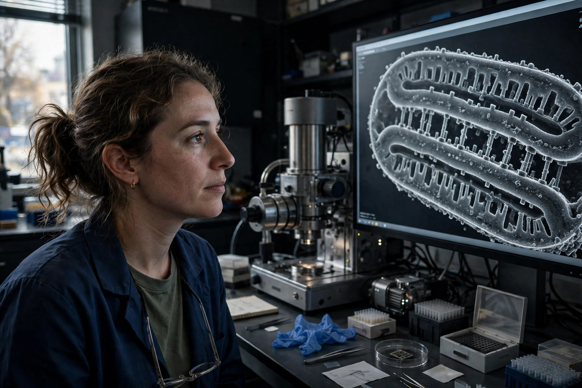
A newly found protein acts as a pillar that sets the space inside mitochondria
Textbook diagrams draw mitochondria with two membranes and a thin gap between them — and until now, no one knew what set the width of that gap. University of Ottawa researchers led by Dr. Luca Pellegrini report in Science Advances (published October 14, 2026; reported October 23, 2026) that a protein called SPRIZ (gene CCDC127) is a rigid tether spanning the outer and inner mitochondrial membranes, locally setting the volume of the intermembrane space. Widening that space by even a few nanometres changed molecular access and slowed mitochondrial calcium uptake, and deleting the gene in mice changed the spacing while leaving overall mitochondrial structure intact.
Read article →
A new form of cell death starts inside mitochondria — and it wakes the immune system up
Cell death is usually quiet. Memorial Sloan Kettering researchers, in work highlighted October 22, 2026, describe a second way the mitochondrial proteins BAK and BAX can assemble: instead of releasing cytochrome c for silent apoptosis, they form a cross-membrane arrangement that lets mitochondrial DNA escape into the cell, triggering inflammation, catastrophic energy failure and a death the team calls baciptosis. It could be induced in human cancer cells with impaired caspase activation — raising the idea of making tumors die loudly enough for the immune system to notice.
Read article →
Cells build a new trash-sorting station inside stressed mitochondria — in lab models
When a mitochondrion is flooded with damaged proteins, the cell does not just throw them away — it sorts them. A Nature study published October 21, 2026 from Nankai University describes SIMPA (stress-induced mitochondrial protein aggregates): under stress, damaged proteins gather into aggregates inside mitochondria in a process that depends on the chaperone GRP75, and are then cleared through contact sites where mitochondria touch the endoplasmic reticulum. The pathway also appeared in mouse liver-injury models, and boosting GRP75 eased liver damage in mice.
Read article →
A mitochondrial RNA switch helps lymphoma cells refuse to die — turning it down restored cell death — in lab models
Diffuse large B-cell lymphoma (DLBCL) is the most common aggressive non-Hodgkin lymphoma, and tumors that resist the antibody drug rituximab have a poor prognosis. A Scientific Reports study published October 20, 2026 identifies a long non-coding RNA, SNHG29, as a resistance mediator that works at the mitochondrion: SNHG29 collaborates with ribosomal proteins and the protein CYLD, keeping phosphorylated CYLD high on mitochondria and apoptosis (programmed cell death) switched off. Down-regulating SNHG29 lowered mitochondrial phosphorylated CYLD and promoted apoptosis in rituximab-resistant DLBCL models, including mouse xenografts.
Read article →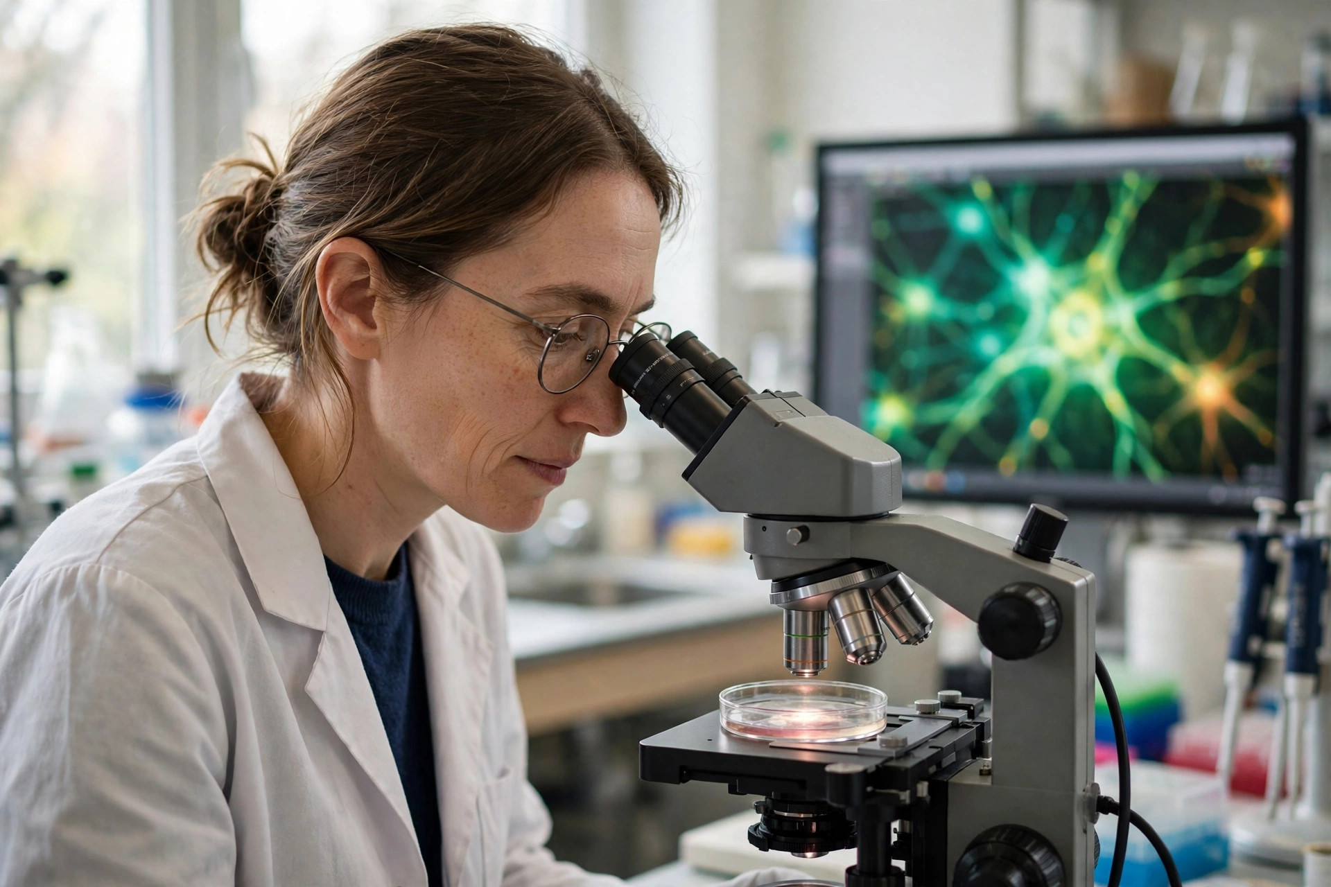
In human neurons, aging does not break the cleanup crew — it breaks the delivery route to the lysosome
Damaged mitochondria are supposed to be recycled by mitophagy, but whether that recycling fails in aging human neurons — not just in mice — was unknown. A Frontiers in Aging Neuroscience study using neurons directly reprogrammed from the skin cells of healthy donors aged 11 to 91 reports the first demonstration of age-related mitophagy impairment in human neurons: in older donors' neurons, mitochondria lost membrane potential and fragmented, then piled up inside autophagosomes and lysosomal vesicles without being eliminated — because their delivery to acidified lysosomes faltered, even though the lysosomes' own enzymes still worked.
Read article →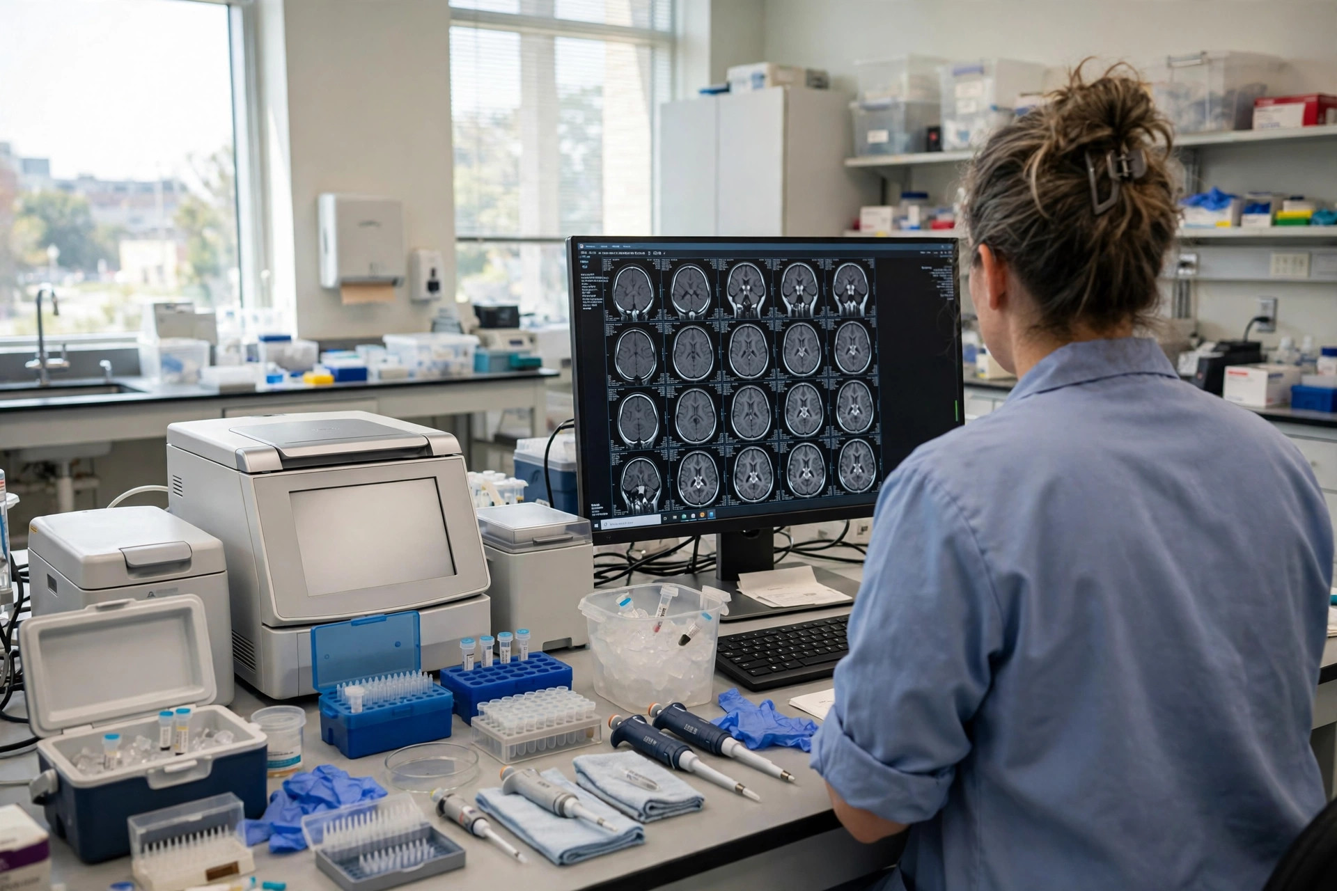
One broken fission gene, a lifetime of seizures: a new DNM1L variant explains a child's refractory epilepsy
Mitochondria must constantly divide to meet the brain's energy demands, and the dividing is done by a protein called DRP1, encoded by the gene DNM1L. A case report published October 19, 2026 in the Orphanet Journal of Rare Diseases describes a previously unreported de novo DNM1L variant (c.1093A>G; p.Arg365Gly) in a 12-year-old girl with severe developmental delay, microcephaly, ataxia and refractory seizures: her neurons showed pronounced mitochondrial hyperfusion — mitochondria stuck together because fission had failed — alongside widespread neuronal loss found after death.
Read article →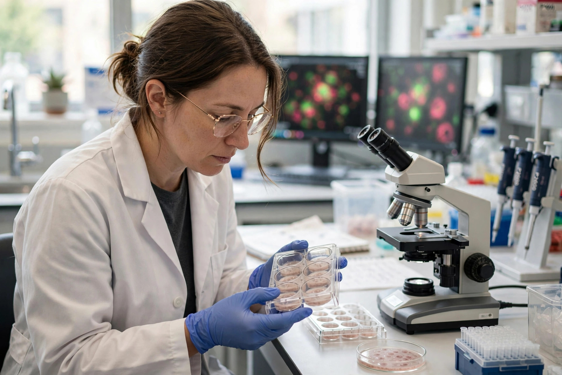
Lung tumors run their mitochondria hot: silencing one complex I gene collapses them — in lab models
Most cancer metabolism stories are about tumors abandoning their mitochondria. A study published October 19, 2026 in npj Precision Oncology reports the opposite for non-small cell lung cancer (NSCLC): the complex I gene NDUFS2 is upregulated in tumors, enriched in malignant cells, and tied to worse clinical features — and silencing it caused profound mitochondrial dysfunction, ATP depletion and loss of viability, migration and invasion in cancer cells, and suppressed xenograft growth in mice. NDUFS2 also switched on the Akt-mTOR growth pathway, making it a candidate drug target — in preclinical models only.
Read article →
A stress enzyme in bone stem cells links diabetes and alcohol to mitochondrial failure and bone loss — in lab models
Diabetes and heavy alcohol use both weaken bone, and a study published October 20, 2026 in Nature Communications points to a shared mitochondrial mechanism: in bone marrow skeletal stem cells, nutrient excess switches on an enzyme called ACSS1, which rewires acetate metabolism, disables a second enzyme (ME2) by acetylation, and traps the cells in a cycle of redox imbalance and mitochondrial dysfunction that blocks bone formation. Deleting ACSS1 — or delivering a non-acetylatable ME2 — restored mitochondrial resilience and bone building in male mouse models of type 2 diabetes and chronic alcohol use.
Read article →
A plant sterol in nuts and seeds disrupts glioblastoma mitochondria — in lab models
Stigmasterol, a cholesterol-like compound found in vegetable oils, legumes, nuts and seeds, suppressed glioblastoma growth in cell culture and mouse xenografts by collapsing mitochondrial function: membrane potential and complex I activity fell while mitochondrial reactive oxygen species rose, in a BMC Cancer study reported October 19, 2026. The tumor's attempted escape — switching on a gene called NDUFA4L2 — also emerged as a drug target. This is preclinical work only; nothing is known about the compound in patients.
Read article →
Why diabetic nerves fail from the feet up: a new review puts damaged mitochondria at the center
Diabetic neuropathy — numbness, burning pain, and silent foot damage — is usually blamed on high blood sugar alone. A comprehensive review published October 17, 2026 in the Journal of Translational Medicine argues the deeper story is mitochondrial: long nerve fibers have extreme energy demands, and when their mitochondria fail, a cascade of oxidative damage, calcium overload, failed cleanup and inflammatory signaling dismantles axons from the far end inward. No disease-modifying therapy exists yet; the review maps where one might come from.
Read article →
A body-clock gene keeps the diabetic heart's mitochondria burning glucose — in lab models
Diabetic cardiomyopathy — heart failure driven by diabetes itself — involves a forced switch away from glucose burning that starves the heart's mitochondria. In Frontiers in Cardiovascular Medicine, researchers report that the circadian gene NPAS2 protects the diabetic mouse heart by preserving the activity of pyruvate dehydrogenase (PDH), the gatekeeper enzyme for glucose oxidation: removing NPAS2 worsened heart damage, and restoring it with a gene-therapy virus improved heart function. The work is in mice and rat heart cells only.
Read article →
A mitochondrial transport protein builds up in the muscle of people with type 2 diabetes — and exercise brings it down
Skeletal muscle does most of the body's glucose disposal, and its failure to respond to insulin is a primary defect in type 2 diabetes. A study published October 19, 2026 in Nature Communications found that Miro1 — a protein on the outer mitochondrial membrane — accumulates in the muscle of patients with obesity and type 2 diabetes but not obesity alone, tracks with worse insulin sensitivity, and falls when patients exercise. Deleting Miro1 from mouse muscle improved mitochondrial respiration and glucose uptake.
Read article →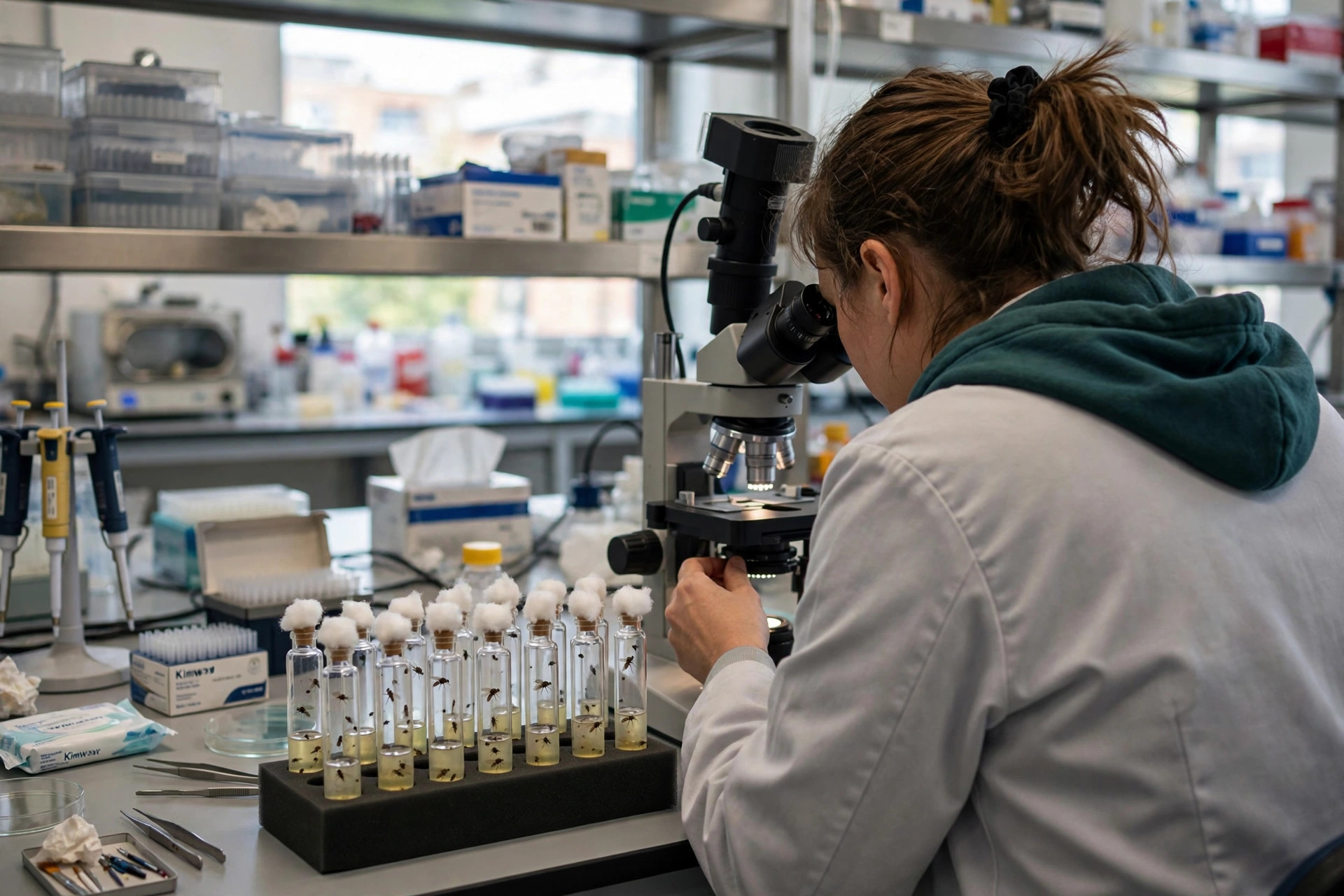
In fruit fly testes, a support cell keeps stem cells going by managing mitochondria in its neighbors
Stem cells do not maintain themselves alone. A Cell Death Discovery study published October 17, 2026 found that the gene Mppa, acting in cyst stem cells of the Drosophila testis, sustains those stem cells and orchestrates germ-cell differentiation by non-autonomously regulating mitochondrial homeostasis — one cell type controlling the mitochondrial health of another. It is basic fly biology, valuable because mitochondrial support between cells is an emerging theme from fertility to transplantation.
Read article →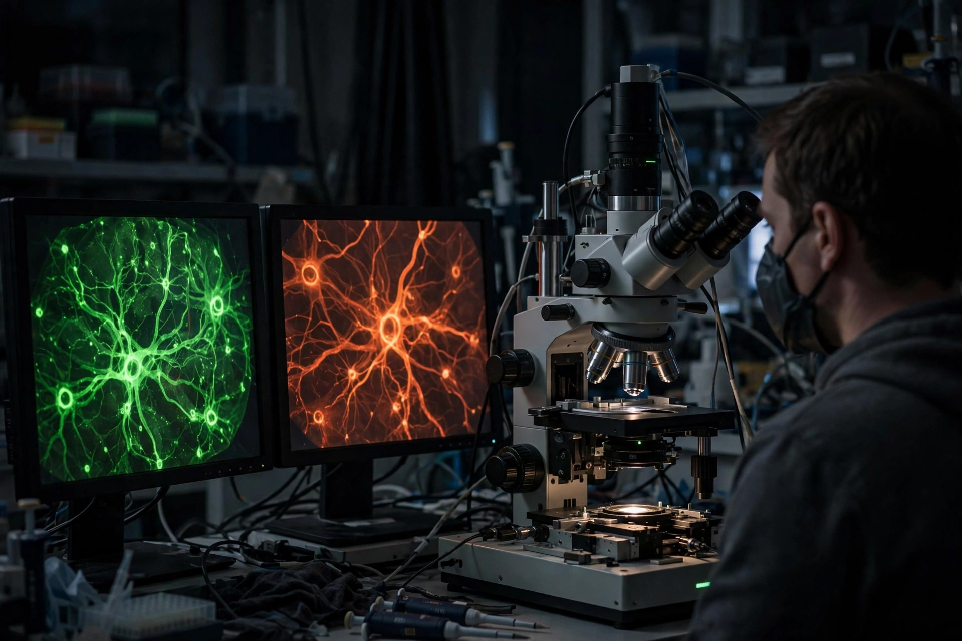
A microscope that watches brain metabolism and oxygen at the same time — in an Alzheimer's mouse model
Mitochondrial dysfunction and poor brain oxygenation are both implicated in Alzheimer's disease, but they are usually measured separately. A Neurophotonics study reported October 17, 2026 used two-photon FLIM-PLIM microscopy to capture metabolic and oxygen information together in living brain tissue from an Alzheimer's mouse model — a framework for seeing how energy failure and oxygen supply change in step as disease develops. Mouse work only; it does not show the same patterns occur in people.
Read article →
A mitochondrial fusion protein has a second job — stopping toxic protein clumps — and its failure may explain a nerve disease
Mitofusin 2 (MFN2) was known for fusing mitochondria together. A Nature Communications study from Cologne, Bochum, Padova and Angers reports that MFN2 also works with the proteasome and molecular chaperones to prevent newly made proteins from aggregating — and that in skin cells from people with Charcot-Marie-Tooth disease, MFN2 mutations disable that protective role, causing damaging protein clumping. The finding reframes how a mitochondrial protein can drive a disease whose other genes have nothing to do with mitochondria.
Read article →
Two mitochondrial quality-control genes stand out in rheumatoid arthritis blood
Rheumatoid arthritis is usually told as an immune story. A Scientific Reports analysis combining bulk and single-cell RNA sequencing from 248 patients and 88 controls found two mitochondrial quality-control genes behaving in opposite directions — CDC42 switched up, RBBP7 switched down — with signatures tied to monocytes, B cells and the JAK-STAT pathway that existing arthritis drugs already target. The findings are associations in blood, not proof of cause.
Read article →
How many copies of mitochondrial DNA a tumor carries tracks with its mutations — and its response to chemotherapy
A multi-cancer analysis in The FEBS Journal (2026) found that mitochondrial DNA copy number correlates with the extent of genetic mutations in cancer cells, carries a signature of compensation for impaired mitochondrial function, and is associated with differing expression of genes linked to chemotherapy response and tumor growth and survival.
Read article →
In Parkinson's, the brainstem neurons that fail first carry heavy mitochondrial DNA damage — and a hint that some fight back
The first single-cell analysis of mitochondrial DNA in vulnerable Parkinson's brainstem neurons, published October 7, 2026 in Brain, found extensive large-scale deletions — concentrated in the major arc that holds key energy genes — alongside a potentially protective response involving the mitochondrial quality-control gene PINK1, most pronounced in people who lived longer after diagnosis.
Read article →
A new imaging library captures mitochondrial cell death frame by frame — and anyone can use it
When a cell commits to die, the decisive step happens at the mitochondrial outer membrane. A dataset published October 15, 2026 in Scientific Data records that moment at scale: mouse embryonic cells treated to alter mitochondrial dynamics, then induced into apoptosis and imaged by high-throughput spinning-disk confocal microscopy — a shared resource for testing how membrane permeabilization really unfolds.
Read article →
Two experimental compounds wreck glioblastoma cells by destroying their mitochondria — in lab models
Glioblastoma is one of the hardest cancers to treat, in part because its cells resist the standard drug temozolomide. A study published October 16, 2026 in Oncogene reports that two novel compounds, EPIC-1027 and EPIC-1042, destroy mitochondrial morphology and function in glioblastoma models by driving p62-mediated degradation of respiratory-chain complex proteins — a strategy aimed squarely at the tumor cell's energy supply.
Read article →
In PCOS, a hidden blood-vessel flaw appeared only when blood sugar rose — in lab-grown cells
Polycystic ovary syndrome carries a higher risk of cardiovascular disease that hormones alone do not explain. Using blood-vessel cells grown from PCOS patients' reprogrammed stem cells, researchers found a mitochondrial defect that stayed invisible until glucose climbed — with the enzyme myeloperoxidase (MPO) as an unexpected accomplice, and a possible target.
Read article →
Mammalian cells make and store elemental sulfur, and it shields them from a rust-like form of cell death
Elemental sulfur was thought to belong to bacteria and hot springs, not human biology. A study in Science reported October 15 that mammalian cells produce cyclo-octasulfur (S8), store it in mitochondria and lipid droplets, and use it to generate antioxidant supersulfides that suppress lipid peroxidation and ferroptosis — the iron-driven cell death linked to tissue injury and aging.
Read article →
A textbook story about how ceramides open death channels in mitochondria did not hold up in new membrane tests
Ceramides — fatty molecules that build up under cellular stress — have long been said to punch channels through the outer mitochondrial membrane to trigger cell death. In a Scientific Reports study published October 15, ceramides alone could not pass even the smallest test markers through model membranes, and could not substitute for cardiolipin in helping the death protein Bax assemble its pores.
Read article →
$12 million federal grant puts mitochondrial energy at the center of brain-aging research
The National Institute on Aging has awarded $12 million over five years to a UTHealth Houston-led team to map how failing mitochondrial energy production shapes memory, mood, and Alzheimer's risk — including blood signatures that might flag brain energy failure years early, and how those pathways differ in women and men.
Read article →
Most people with mitochondrial disease were still alive a decade after diagnosis, a national study of 1,107 patients found
Survival in mitochondrial disease has long been guesswork drawn from small clinic cohorts. A nationwide English study in Genome Medicine linked genetic diagnoses to national health records for 1,107 people and found 80.3% were alive ten years after diagnosis — and no higher cancer rate than the general population.
Read article →
A clearer way to watch mitochondria inside hard-to-see nerve-support cells — in mice
Terminal Schwann cells — the support cells capping the junction where nerves meet muscle — have been nearly impossible to study in place. A methods paper in the Journal of Visualized Experiments shows how to measure their mitochondrial shape and organization inside intact mouse tissue, and already picks up differences between healthy and dystrophic muscle.
Read article →
Six weeks of heavy alcohol rewired the heart's mitochondrial RNA machinery — in rats
Chronic heavy drinking is a leading cause of dilated cardiomyopathy, and mitochondrial damage is part of the mechanism. In a Frontiers in Cardiovascular Medicine study, rats fed an alcohol diet for six weeks showed hundreds of gene changes in the heart's left ventricle — with the pathways that process and degrade mitochondrial RNA switched up, and daily-clock and immune-defense pathways switched down.
Read article →
A fuel-ferrying mitochondrial gene emerged as a causal player in Parkinson's — in data models
Low oxygen and failing mitochondrial energy production are hallmarks of Parkinson's disease, but the connecting regulators have been unclear. A multi-omics analysis published October 13 in npj Parkinson's Disease built a seven-gene diagnostic model and, using genetic causal inference, singled out MPC2 — a carrier that ferries fuel into mitochondria — as depleted in the dopamine neurons that die in Parkinson's.
Read article →
Deleting a heart-rhythm and diabetes gene disrupted mitochondria and insulin signals in lab-grown human neurons
KCNQ1 is known for heart-rhythm disorders and type 2 diabetes risk. In a Communications Biology study published October 13, deleting the gene from human stem cells slowed early neuron growth and disturbed mitochondrial function and insulin signaling — a clue to why the same gene keeps appearing across heart, metabolic, and brain conditions.
Read article →
Blocking mitochondria's cleanup system made a standard leukemia drug work harder — in lab and mouse models
Acute myeloid leukemia cells lean on mitophagy — the recycling of worn-out mitochondria — to survive. A study published October 13 in Leukemia found that disabling the mitophagy regulators PINK1 or PHB2 made leukemia cells, including cells from 20 patients, far easier for the standard drug venetoclax to kill, in dishes and in mouse xenografts.
Read article →
Younger astronauts showed the strongest mitochondrial stress signals in orbit
Blood samples from 10 International Space Station crew members, collected between 75 and 210 days into their missions, showed an age-dependent response to spaceflight — and, counterintuitively, the younger astronauts showed more pronounced gene changes and more severe signs of mitochondrial dysfunction.
Read article →
Damaged mitochondrial DNA marked the leukemia cells that answered imatinib better
In 120 people with chronic myeloid leukemia, cancer cells carrying more mitochondrial DNA damage relied less on mitochondrial energy production — and responded better to the frontline drug imatinib. Cells with fitter, more active mitochondria held out longer, pointing to a new way to spot harder-to-treat disease.
Read article →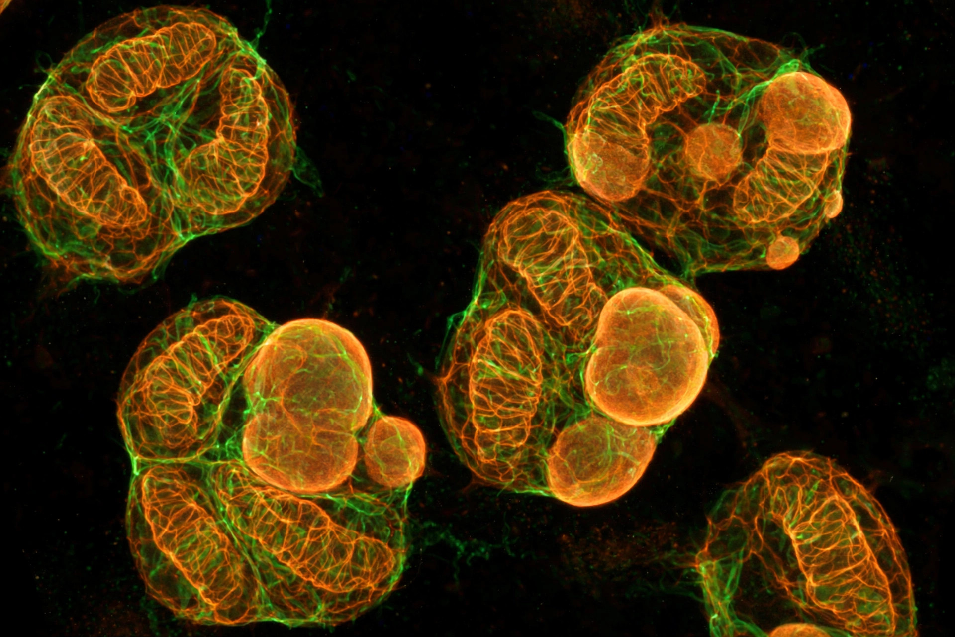
A tumor protein wrecks neutrophils' mitochondria to feed leaky blood vessels in bladder cancer — in lab models
Researchers found that the secreted tumor protein LRG1 sabotages the mitochondria of neutrophils, driving them to cast DNA "nets" that build leaky, chaotic tumor blood vessels — and blocking LRG1 made chemotherapy and immunotherapy work better in mice.
Read article →
Review: failing mitochondria may be the organizing hub of aging inflammation
A new GeroScience review argues that mitochondrial bioenergetic failure, cellular senescence, and sterile inflammation converge through mitochondria — and soberly notes where the evidence, including for urolithin A, falls short.
Read article →
A little-known protein guards brain mitochondria after bleeding stroke — in mice
Chongqing Medical University researchers identified TSPOAP1 as a previously unrecognized protector of the mitochondrial membrane pore in neurons after intracerebral hemorrhage — losing it spilled cytochrome c and drove brain cells to self-destruct.
Read article →
Nine new maps of mitochondrial proteins could unlock drugs for malaria-like infections
Across nine Cell papers, teams from Harvard, the Broad Institute, and Mass General Brigham catalogued the mitochondrial proteins of parasites including Babesia — 525 proteins, the most comprehensive such inventory for its group — offering new drug targets for babesiosis, malaria, and toxoplasmosis.
Read article →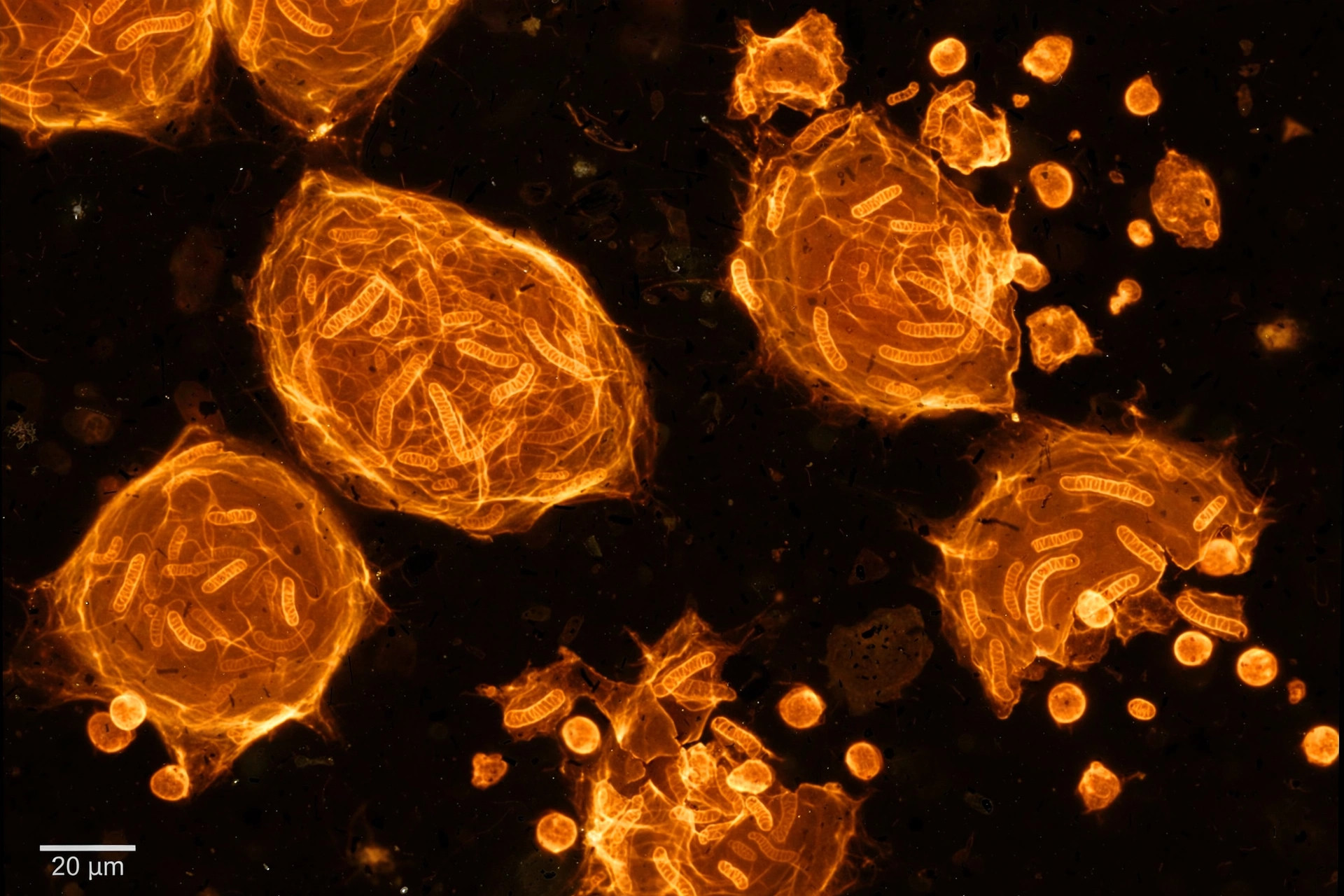
A mitochondria-targeting compound clears chemo-worn cancer cells — in lab models
Researchers at Harbin Medical University and Harvard Medical School found that SMIP004, a small mitochondria-targeting compound, promotes the breakdown of a protein (PDHA) that keeps therapy-induced senescent tumor cells alive — killing them selectively through an energy crisis, and improving chemotherapy outcomes in mice.
Read article →
In ALS, the brain's support cells may be starving motor neurons — in lab models
Using patient-derived stem cells, researchers showed astrocytes carrying the C9ORF72 mutation directly impair mitochondrial transport in motor neuron axons — and boosting the astrocytes' mitochondrial metabolism reversed the deficit.
Read article →
At 194, the world's oldest land animal kept its mitochondrial genes young
A Science Advances study of Jonathan, the 194-year-old Aldabra tortoise, found 272 mitochondrial-function genes with promoters as youthful as a 5-year-old's — while 91-year-old tortoises' looked their age.
Read article →
Transplanting mitochondria could be medicine's future — but first, the field has to solve isolation
A new review in Frontiers in Physiology argues that mitochondrial transplantation — moving healthy mitochondria into damaged tissue — can't scale until the field solves the unglamorous problem first: isolating viable mitochondria without damaging them.
Read article →
Your mitochondria's DNA count may reveal how aggressive a tumor is — and how it will respond to chemo
A multi-cancer analysis in The FEBS Journal ties mitochondrial DNA copy number to tumor mutational load, tumor growth genes, and chemotherapy-response genes — positioning mtDNA as a possible guide to cancer management.
Read article →
Scientists mapped mitochondrial damage in Parkinson's most vulnerable neurons — and found the brain fighting back
In Brain, researchers report the first single-cell look at mitochondrial DNA in the brainstem neurons Parkinson's attacks first — and found a quality-control gene, PINK1, rising to meet the damage, especially in longer survivors.
Read article →
A sabotaged mitochondrial cleanup chain drives blinding retinal scarring — and blocking it worked in mice
In GeroScience, researchers trace the blinding scarring of macular degeneration to a sabotaged mitophagy chain: excess LAPTM5 destroys the ligase that tags broken mitochondria, their DNA leaks, and the immune system fires — but blocking the cascade reversed scarring in aged mice.
Read article →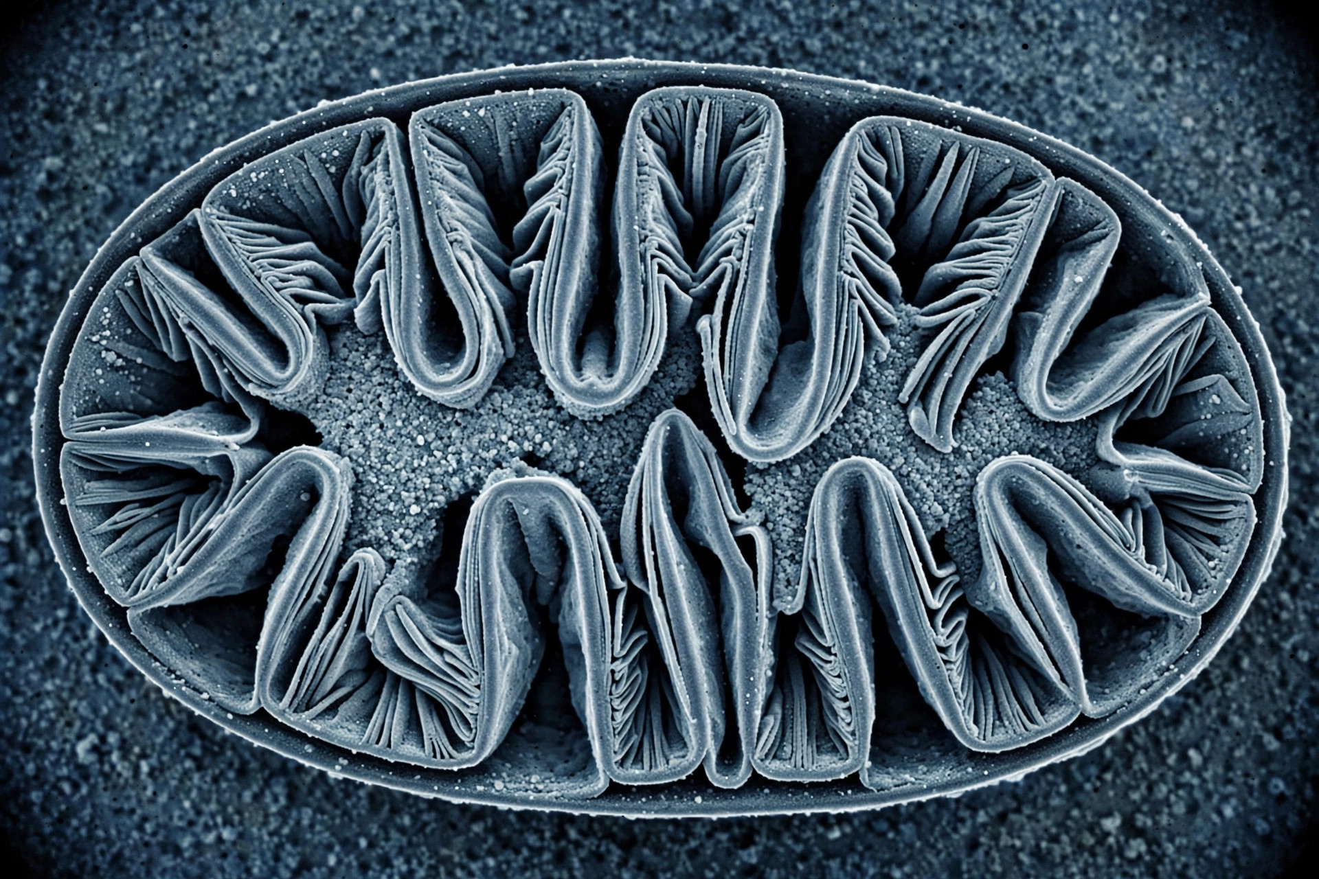
Scientists found the flexible gate that controls your mitochondria's inner folds
Using computer simulations plus a new experimental method, researchers mapped how the Mic60-Mic19 protein pair forms a flexible gate at the mitochondrial cristae — and showed a disease-linked mutation distorts it.
Read article →
The amino acid leucine turns up mitochondrial power by slowing protein cleanup — in cells and roundworms
Cologne researchers found leucine dials down SEL1L, a quality-control protein — leaving more energy-making machinery in place. The lead scientist warns the same trick could backfire, since that cleanup also removes damaged proteins.
Read article →
A mitochondrial protein called NDUFAF2 drives deadly aortic aneurysms — in mice
In iScience, researchers found NDUFAF2 — a complex I assembly factor — upregulated in aneurysm-model mouse aortas; switching it off stabilized vessel walls by restoring mitochondrial fusion and cutting ROS-driven cell death.
Read article →
SLC25A34: the mitochondrial transporter tying your body clock, the cold, and what you eat to fat burning — in mice and cells, with human clues
In Science, researchers identified SLC25A34, a mitochondrial carrier in brown fat controlled by a circadian clock protein, cold exposure, and dietary fat — silencing it cut fuel burning in human brown fat cells, and people with more of it in their fat tended to be leaner.
Read article →
Transplanted mitochondria reboot the aging heart's broken cleanup system — in mice
A new Aging Cell study found that injecting healthy mitochondria into the blood of aged mice restored heart function and cleared a “traffic jam” of half-digested mitochondria — by quieting an energy-sensing HIF-3α→BNIP3 axis that floods aging hearts with cleanup receptors when ATP falls.
Read article →
DUSP4: the enzyme switch that tells fat cells to build mitochondria and burn heat — in mice and cells
Researchers have identified DUSP4 as the controller of a new DUSP4–CRTC3–UCP1 pathway that drives mitochondrial biogenesis genes and calorie-burning thermogenesis in fat — a potential future target in obesity.
Read article →
Microplastics ride the food chain through the body — and at the brain's gate, failing mitochondria open the door — in lab models
A new Nature-portfolio study traced food-chain microplastics through mice and found damage across the liver, kidney and lungs; laboratory work it cites shows microplastics can impair the brain's blood-vessel cells by triggering mitochondrial dysfunction.
Read article →
A food-poisoning bacterium's toxin attacks mitochondria in wounds — in lab and mouse models
The NheABC toxin of Bacillus cereus — best known for food poisoning — punches holes in cell membranes and damages mitochondria in wounded tissue, with the mitochondrial lipid cardiolipin amplifying the damage, researchers at Umeå University report.
Read article →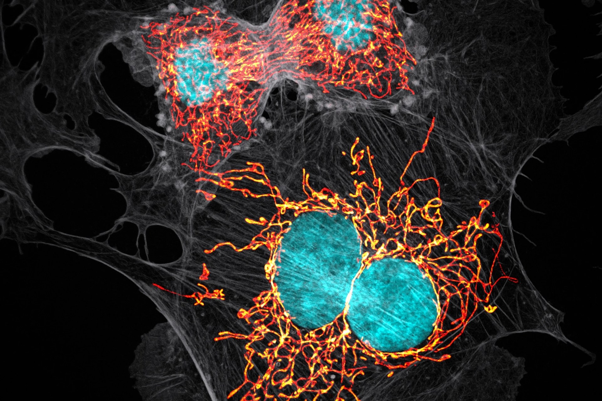
Scientists map how cells flag damaged mitochondria for cleanup — in lab models
RAB1A and RAB1B proteins link the autophagy adaptor OPTN to membrane-building ATG9A vesicles, showing how cells mark the spot where a damaged mitochondrion gets wrapped for recycling, according to a new study from the Tokyo Metropolitan Institute of Medical Science.
Read article →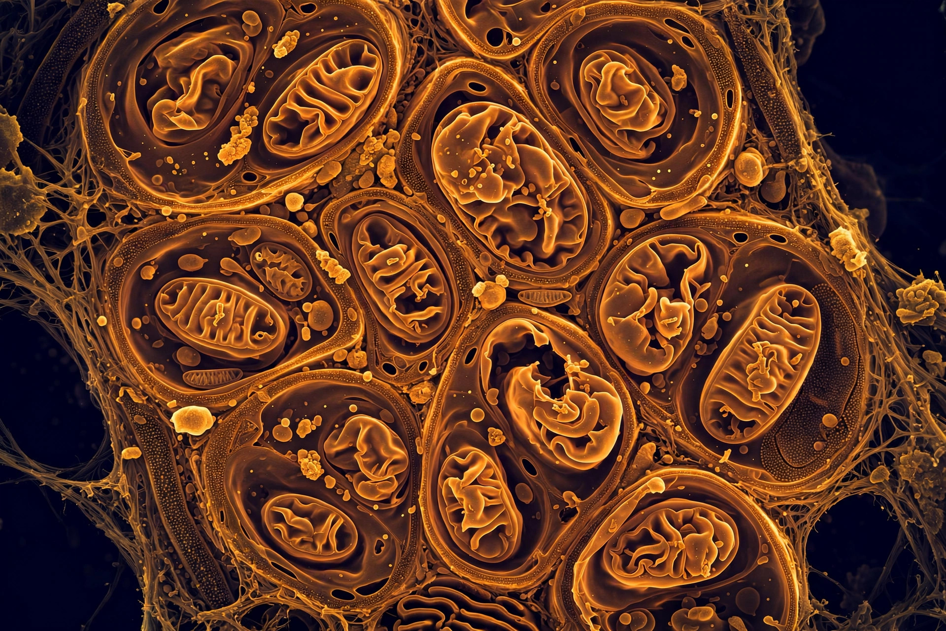
When mitochondria lose their inner folds, liver disease can follow — in lab models
In Cell Death & Disease, Alexander Becker and colleagues show that MIC13-linked disruption of mitochondrial cristae — the inner-membrane folds where energy is made — triggers metabolic failure and early fibrotic remodeling in a liver disease model.
Read article →
Exercise rewires your liver's mitochondria — differently in men and women, in rats
University of Missouri researchers found endurance exercise remodels the mitochondrial Krebs cycle in liver cells through distinct pathways in male and female rats — females built more energy-cycle proteins, males remodeled existing ones.
Read article →
A census of mitochondria across the tree of life finds half their proteins are still mysteries
A Cell study catalogs the mitochondrial protein inventories of plants, parasites, and humans — finding that Leishmania and Trypanosoma carry mitochondrial proteomes 50% larger than ours, and that more than half of all these proteins have unknown functions. All the data is open at mitocarta.org.
Read article →
Chi and Mitochondria: What an Ancient Idea Gets Right — and Where the Evidence Stops
More than two thousand years before anyone knew mitochondria existed, traditional Chinese medicine described a vital energy flowing through the body. Modern science has put that claim to the test. Here is what the published research actually shows — graded against the evidence.
Read article →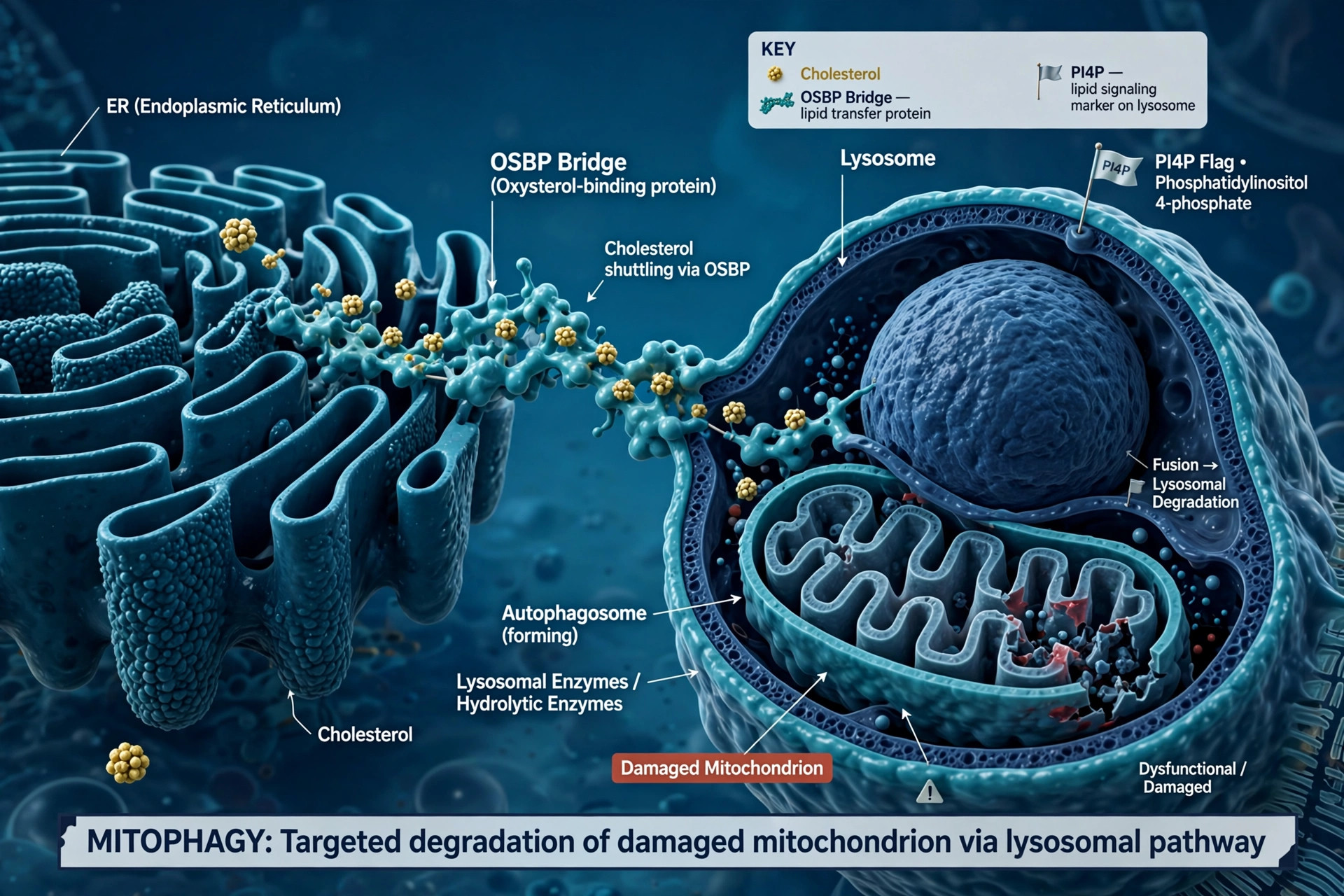
Cholesterol is the supply chain that keeps cellular recycling running — in lab models
When damaged mitochondria land in lysosomes, cholesterol rushes in to armor the recycler's membrane. A Nature Communications study maps the PI4P–OSBP supply chain — and why its failure may matter in Parkinson's and Alzheimer's.
Read article →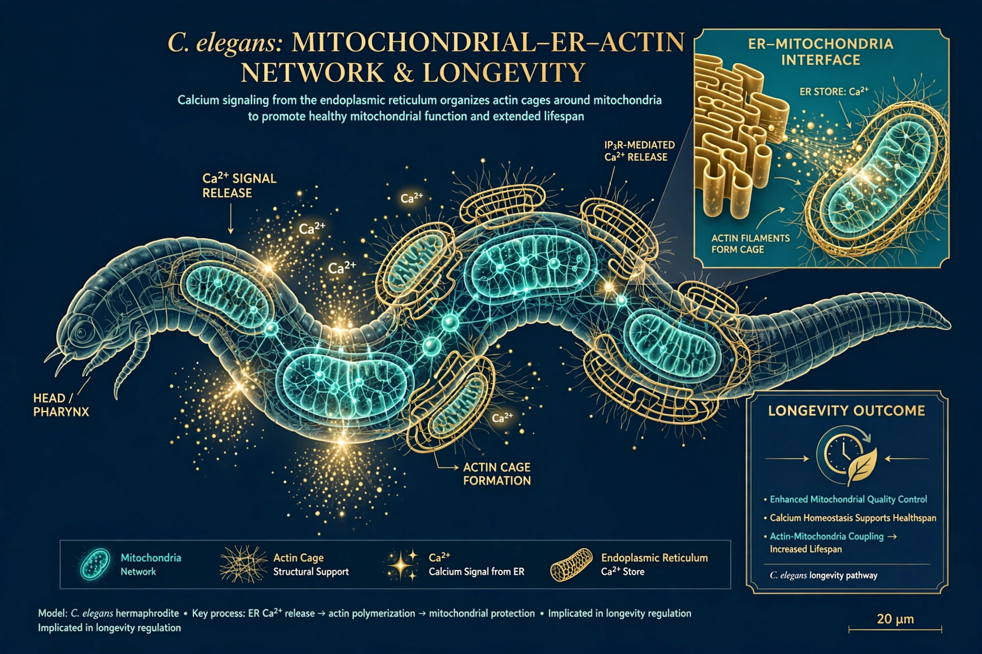
Longer life despite broken mitochondria: worms build calcium-triggered cages — in lab models
In C. elegans, mild mitochondrial stress extends life — but only if ER calcium signaling builds actin 'cages' that keep mitochondria in check. The pathway links two failures that co-occur in aging brain disease.
Read article →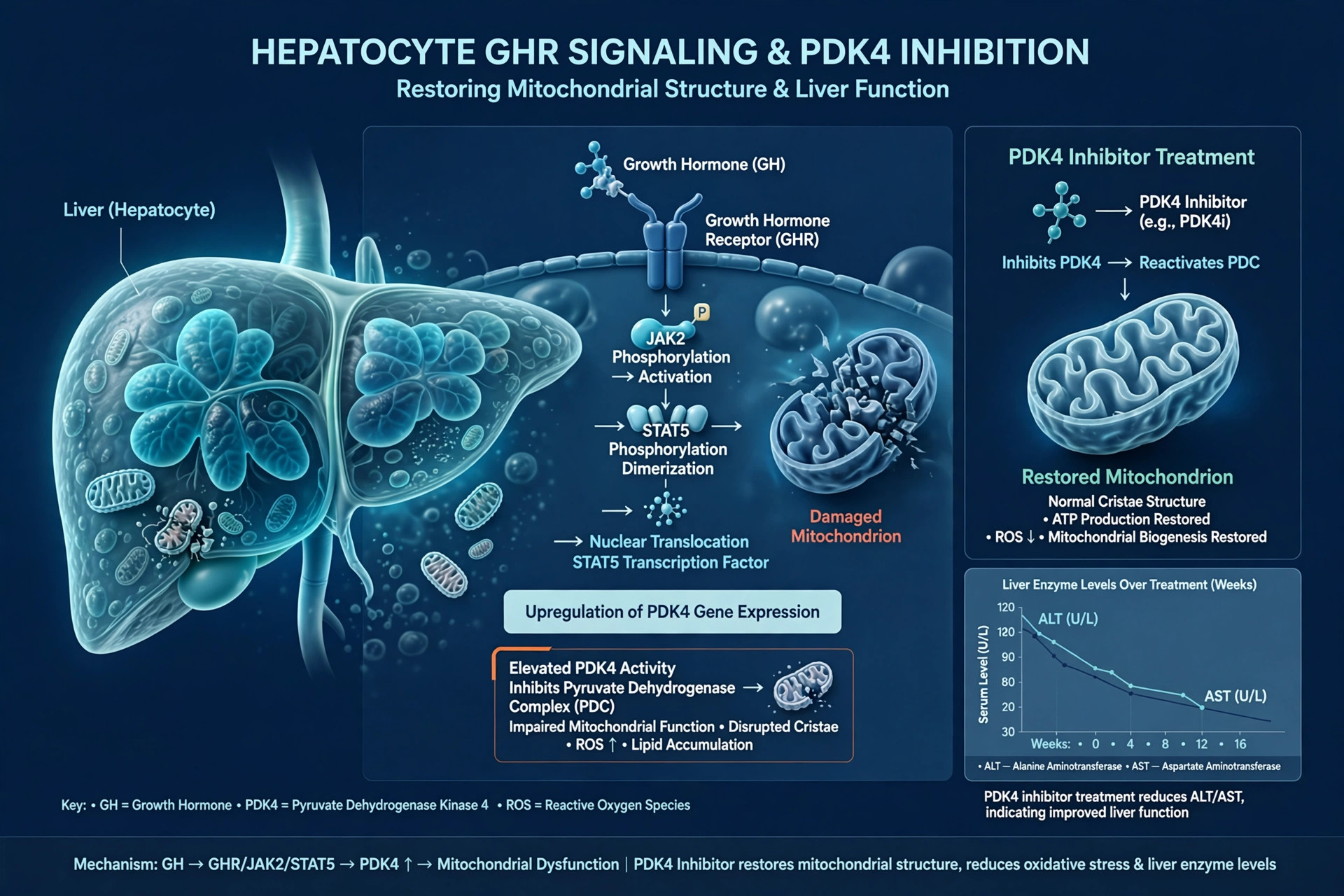
Deleting a liver hormone receptor speeds aging — and blocking PDK4 reverses it, in mice
Knocking out the growth hormone receptor only in liver cells produced fatty liver, mitochondrial failure, and early aging in mice; a PDK4 inhibitor restored mitochondrial structure and cut inflammation.
Read article →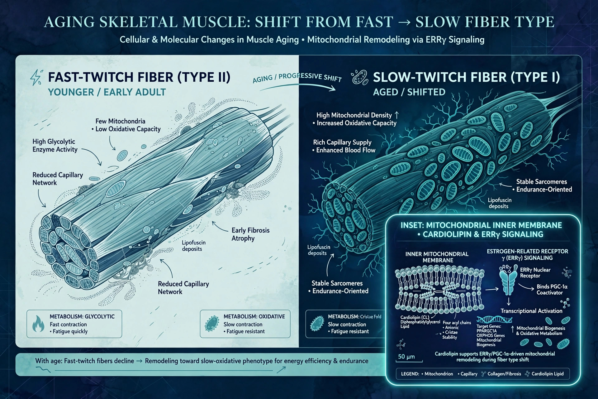
A membrane lipid is the switch for aging muscle — in lab models
A Nature study finds the mitochondrial membrane lipid cardiolipin, acting through estrogen-related receptor γ, controls whether aging muscle keeps its fast fibers or shifts to slow — a new lever on sarcopenia.
Read article →
Light First, Breath Always: Quantum Tunneling, Mitochondria, and the Bible
Quantum tunneling inside your mitochondria is real physics. Surprisingly, some of the Bible's oldest images — light first, a fire that never goes out, breath as life itself — echo it. Here is where the resonance is real, and where it stops.
Read article →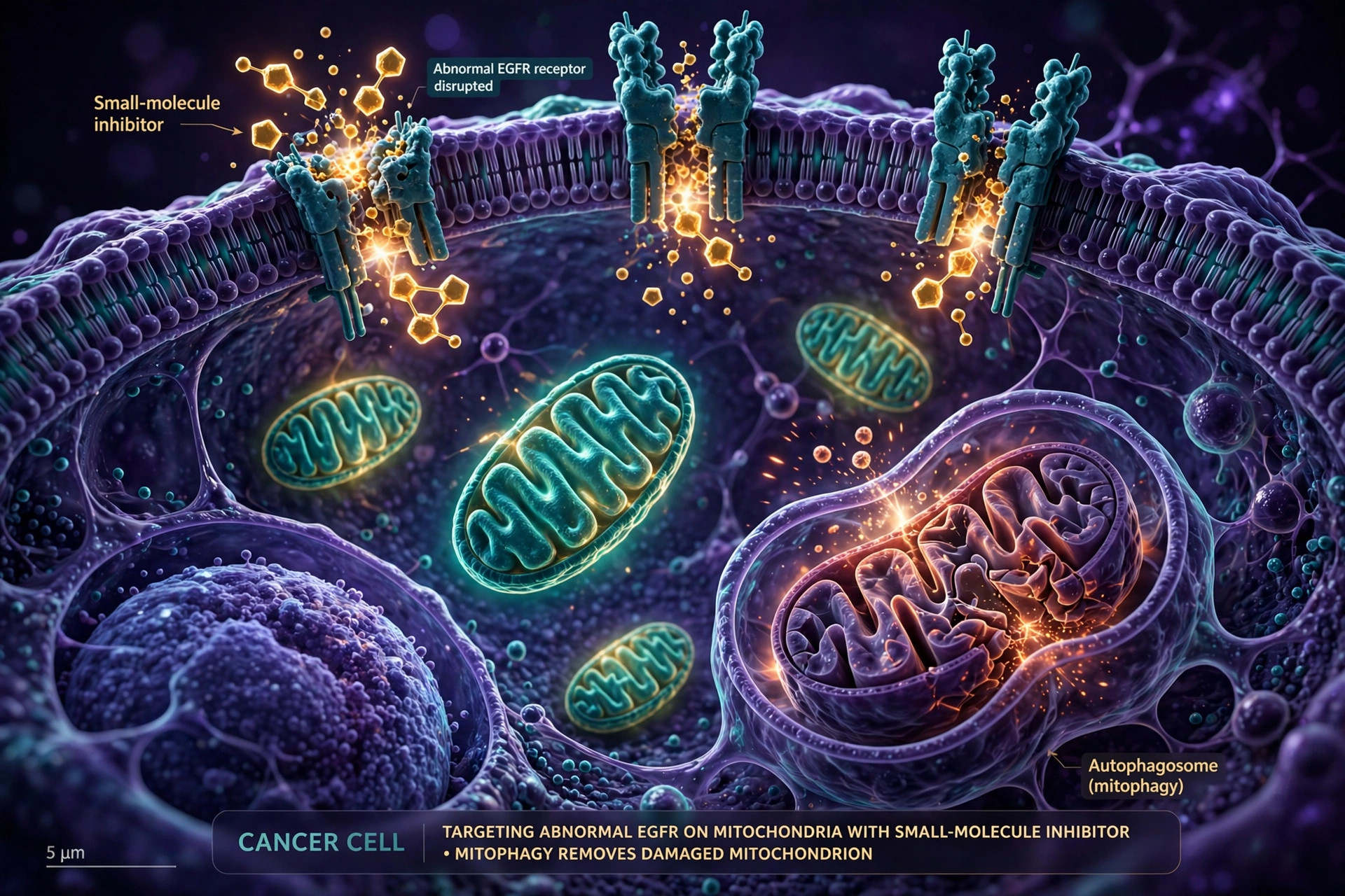
A soap-like molecule turns the tables on breast cancer's mitochondrial trick
Triple-negative breast cancer hides EGFR inside mitochondria to drive growth; screening found cetrimonium bromide degrades it, triggering mitophagy and blocking tumor spread in lab models.
Read article →
Mitochondria in armor: a molecular coating that leaves energy production intact
Researchers in Milan coated mitochondria with metal-organic frameworks at tiny concentrations without harming their function — a step toward armored mitochondria built for transplantation.
Read article →
Exercise's anti-aging effect runs through your mitochondria — and a new study proves the link
A PNAS study found exercise's anti-aging benefits in aged mice and frail adults (average age 78) came from remodeling mitochondria in muscle — and genetically blocking that remodeling erased the benefits, proving causation.
Read article →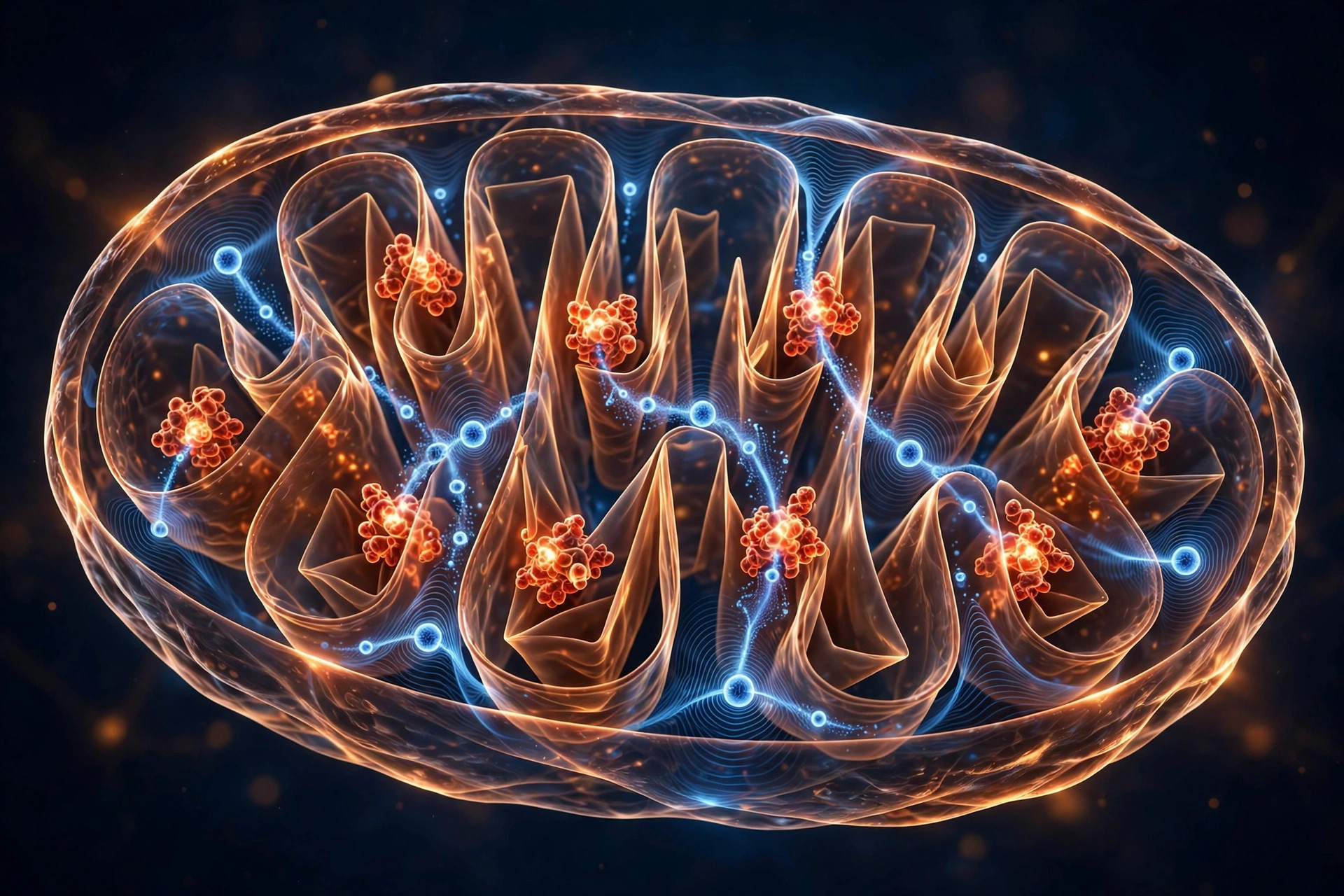
The Quantum Secret Inside Your Mitochondria
Your mitochondria don't just burn fuel — they use quantum tunneling to move electrons. Here's the real, textbook-established science, where it ends, and where the wonder honestly begins.
Read article →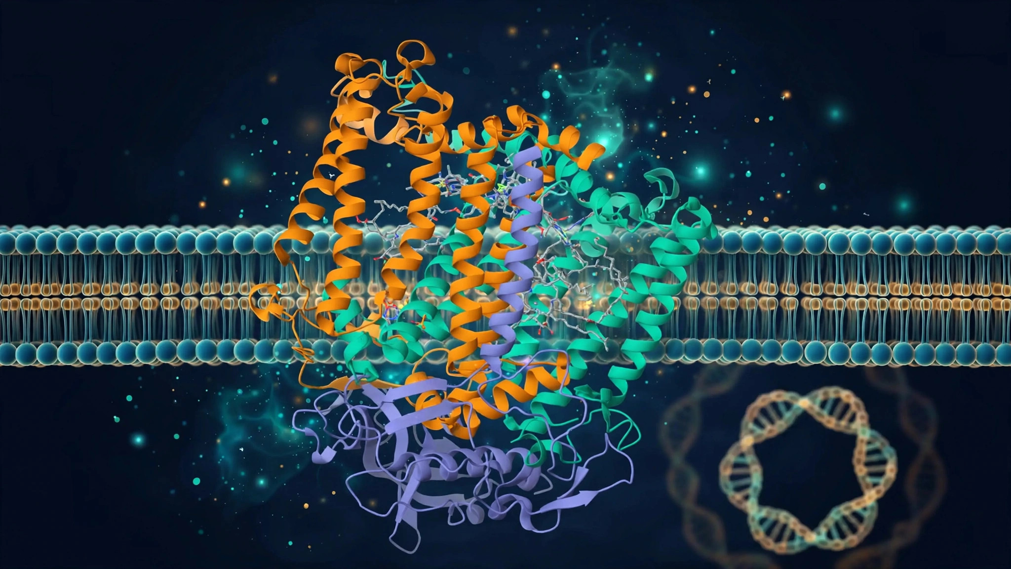
A new map of MT-CYB disease: when mitochondrial complex III fails
A new Frontiers in Neuroscience review synthesizes the full clinical spectrum of MT-CYB-associated mitochondrial disease — a reference for when to suspect complex III disorders and what genetic testing can reveal.
Read article →
Walk more, carry more mitochondria: light activity tied to higher mtDNA copy number
In 256 middle-aged adults, habitual light activity like walking was associated with higher leukocyte mitochondrial DNA copy number; sedentary time and vigorous exercise showed no link.
Read article →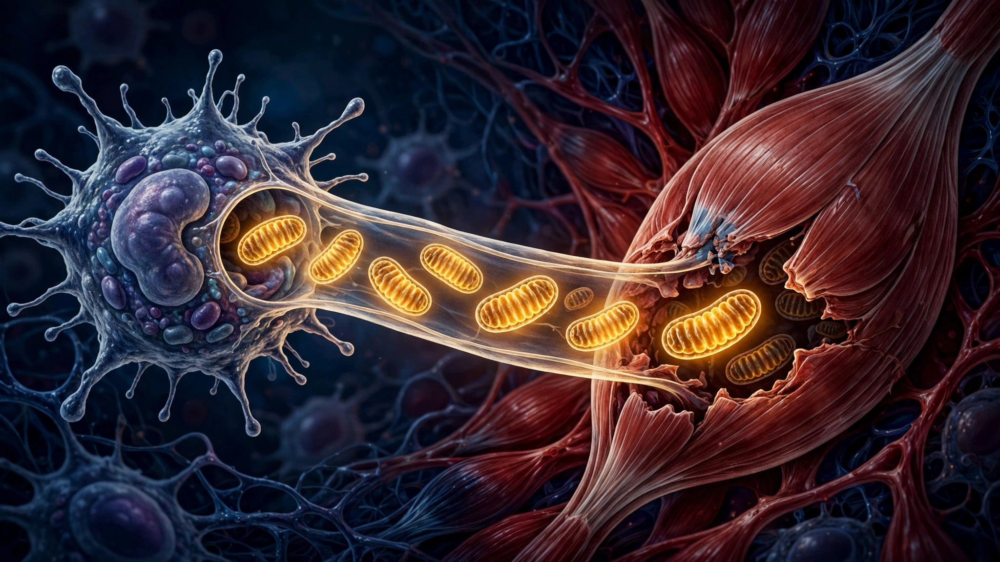
Immune cells donate healthy mitochondria to rescue a failing heart
Activating the Flt3 receptor on macrophages prompted the immune cells to transfer healthy mitochondria into injured heart-muscle cells after a heart attack in animal models, reducing harmful remodeling.
Read article →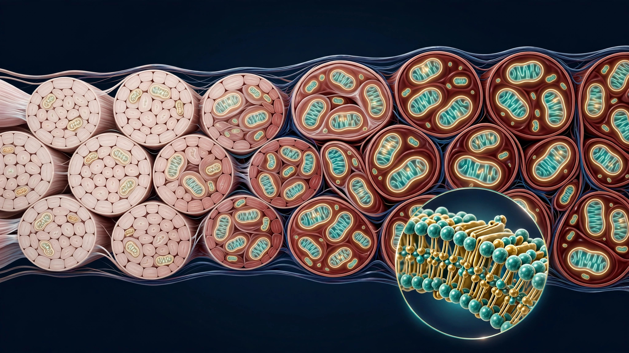
The hidden switch behind an aging-muscle paradox: a mitochondrial fat holds the answer
A new Nature Aging study finds that age-related decline in cardiolipin — a fat found almost only in mitochondrial membranes — drives aging muscle's paradoxical shift to slow-twitch fibers, and partially restoring it reversed wasting in mice.
Read article →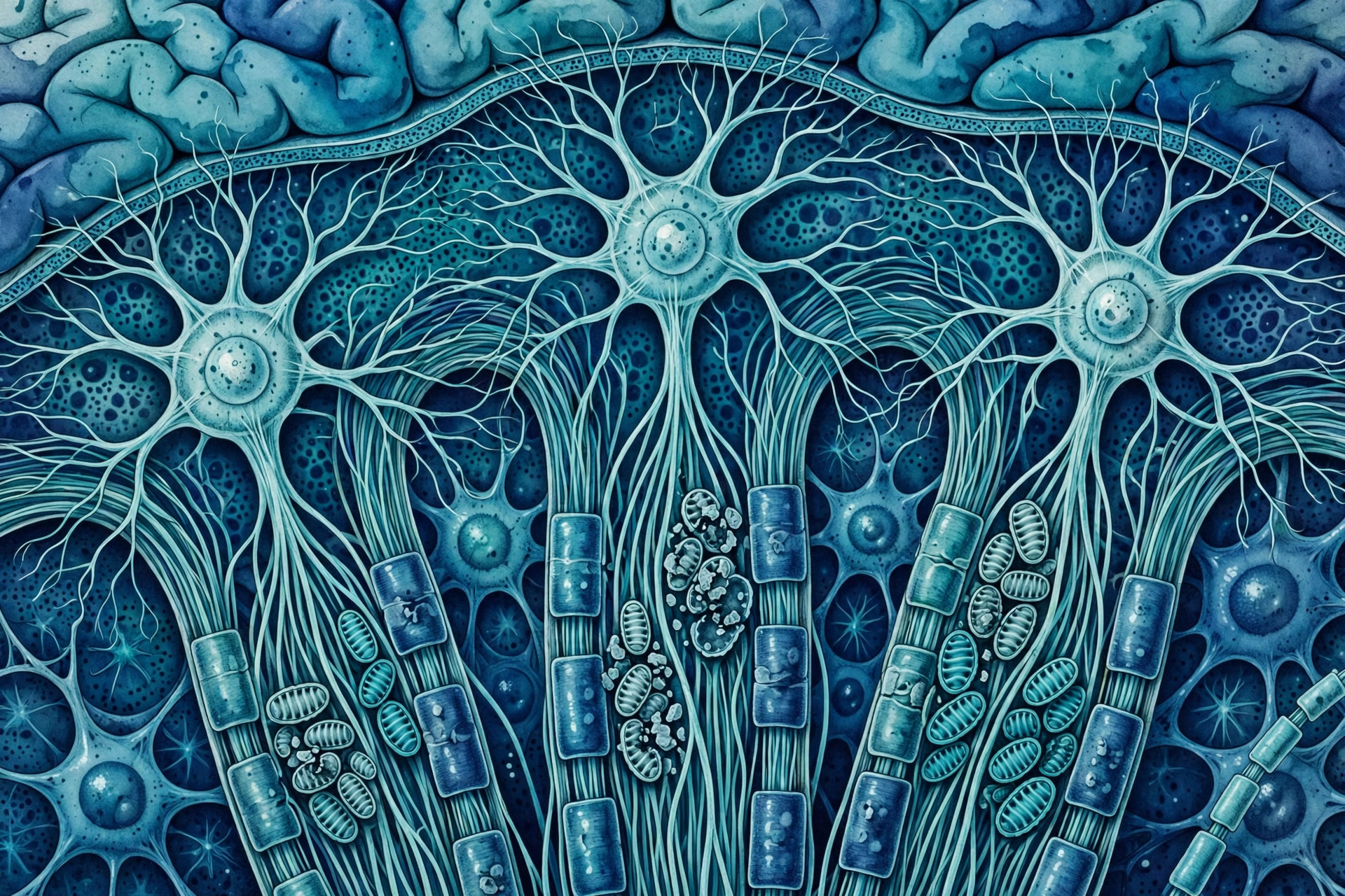
“Mitochondrial plaques” found in Alzheimer’s brains
University of Minnesota researchers identified clustered, damaged mitochondria inside nerve fibers in Alzheimer's brains — suggesting early lysosome failure may precede amyloid plaques.
Read article →
One amino-acid trim keeps mitochondrial complexes from falling apart
Heidelberg and Freiburg researchers found the enzyme ICP55 essential for stabilizing HSP60 and respiratory-chain complexes. A new mechanistic starting point for mitochondrial disease.
Read article →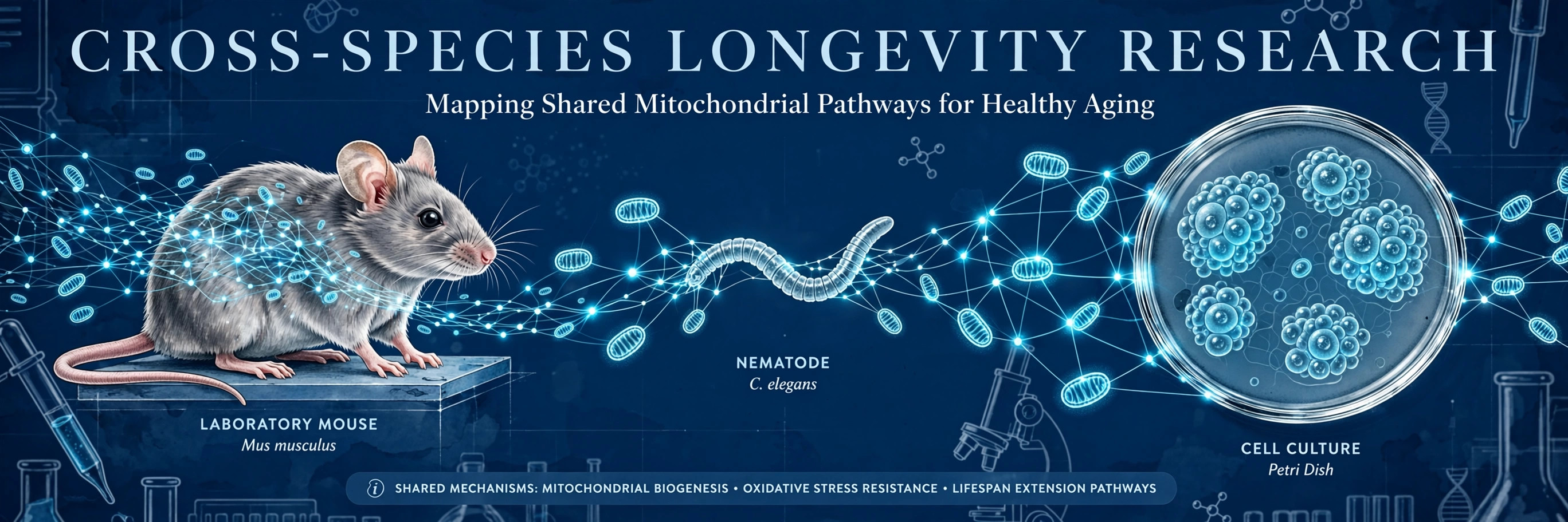
The hits don't always hit: rapamycin and metformin fail in killifish
A rigorous cross-species geroprotector screen across yeast, worms, flies and killifish reports notable negative results: rapamycin and metformin failed in killifish; metformin and resveratrol failed in mice.
Read article →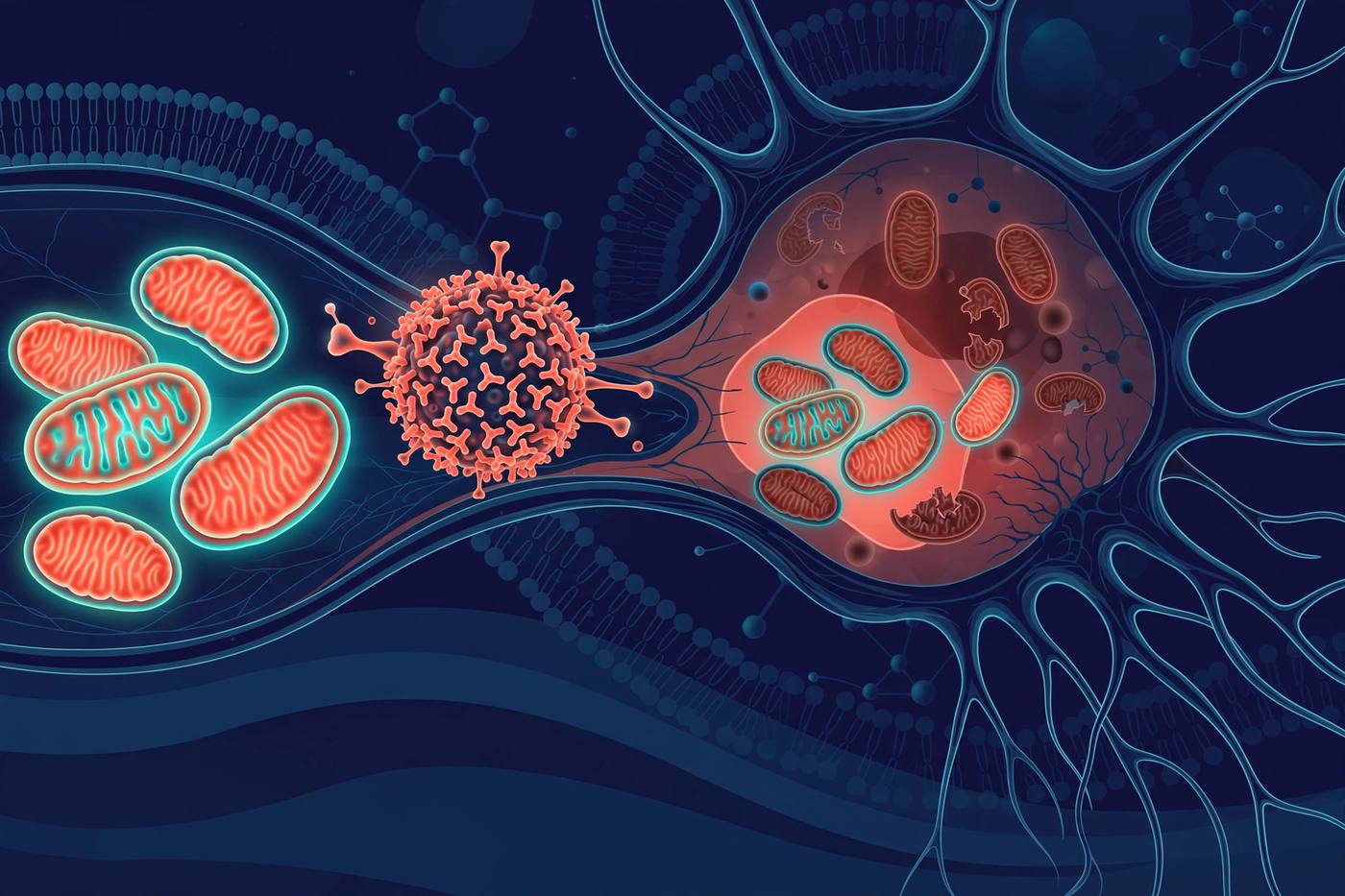
MitoCatch targets precision mitochondrial delivery
A protein-binder system directed donor mitochondria to diseased cells in preclinical models; human testing is still ahead.
Read article →
Masked donor mitochondria in diseased mice
A molecular disguise helped transplanted mitochondria enter cells with defective organelles in a mouse study.
Read article →
Eight babies born after mitochondrial donation in the UK
Published outcomes from mitochondrial donation in the UK found healthy births while long-term risks remain open.
Read article →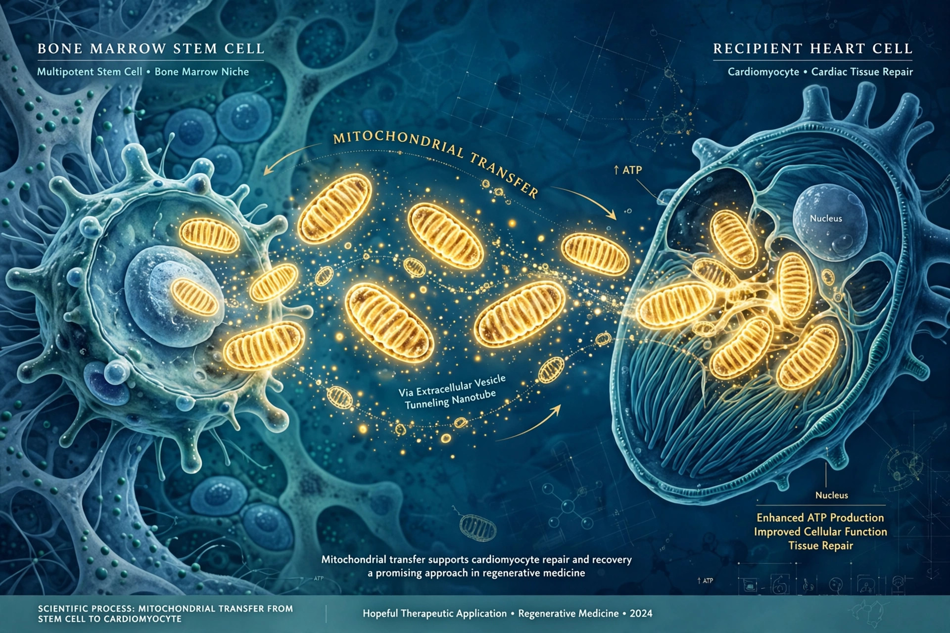
Bone marrow cells deliver mitochondria to heart and brain
A Friedreich’s ataxia mouse model suggests donor-derived immune cells can transfer healthy mitochondria to failing cells.
Read article →
Mitochondrial transplantation enters human testing
Small studies in cardiac shock, stroke and inflammatory myopathy report early safety signals but do not yet prove efficacy.
Read article →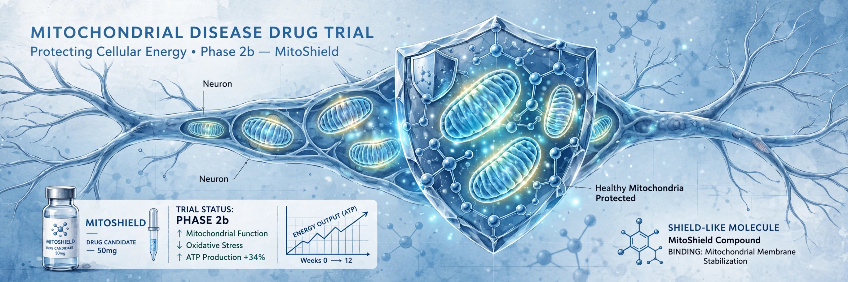
NRG5051 doses first volunteers
A Phase 1 program testing safety and tolerability of a mitochondrial candidate in healthy adults.
Read article →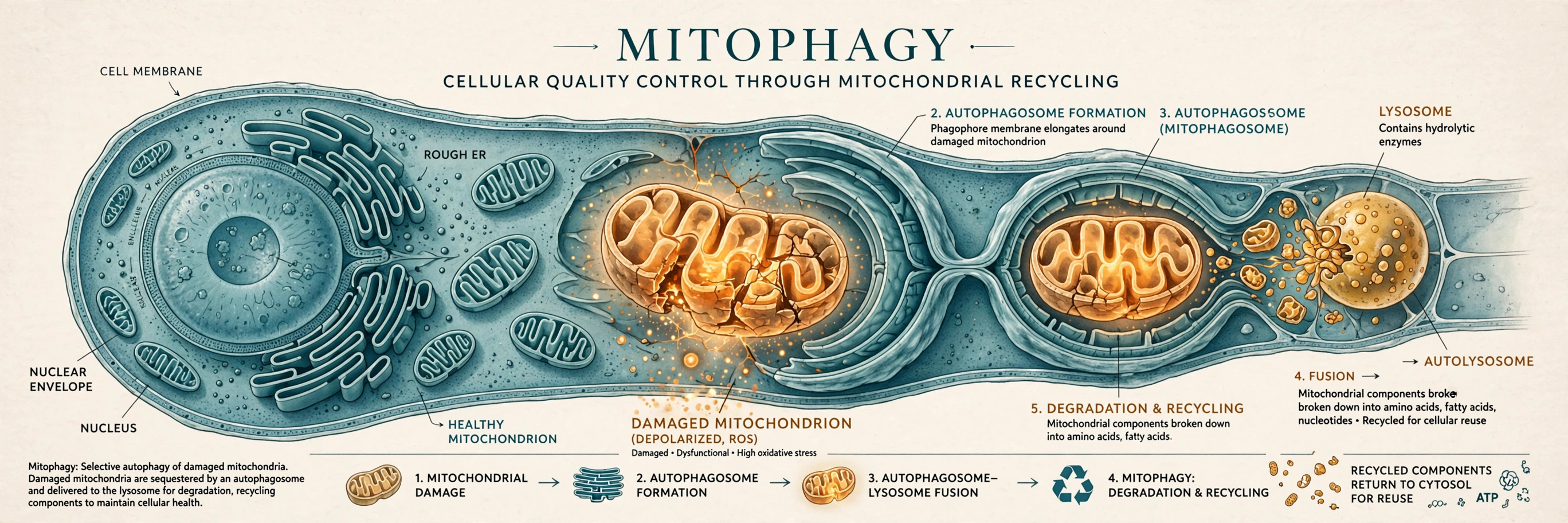
MTX325 targets damaged-mitochondria clearance
The Phase 1b USP30 inhibitor program is designed to enhance mitophagy in Parkinson’s disease.
Read article →
Mitochondrial transplantation research for Duchenne
A 2026 grant supports preclinical mitochondrial transplantation work for Duchenne muscular dystrophy.
Read article →