
Testing
Mitochondrial testing covers the clinical, research, and consumer tests used to measure how well your mitochondria work — from mtDNA sequencing and metabolite panels to full validated workups. Our testing desk examines each method for analytical and clinical validity, so you can judge what a result actually means for your health.

For mitochondrial diabetes, a urine sample found what a blood test can miss
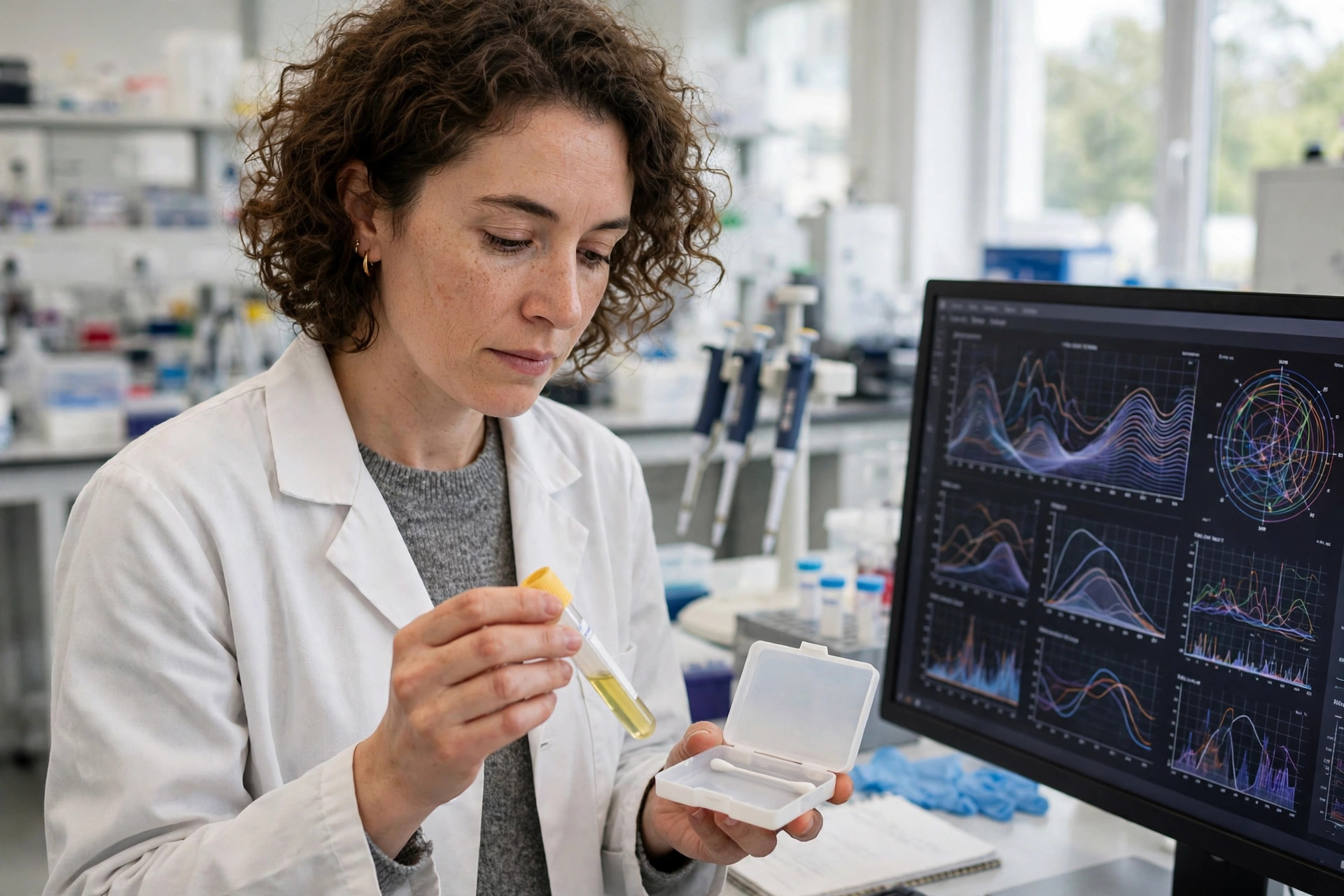
Mitochondrial diabetes is a rare, maternally inherited form of diabetes that is easy to mistake for type 1 or type 2. A study in Frontiers in Endocrinology (Skoczylas and colleagues; published November 11, 2026) sequenced mitochondrial DNA in 496 Polish patients referred with suspected monogenic diabetes after negative nuclear DNA testing, and found pathogenic mtDNA variants in 21 (a diagnostic yield of 4.23%), most often m.3243A>G (17 of 21). In 30 m.3243A>G carriers, the share of mutated mitochondrial DNA (heteroplasmy) followed a clear tissue hierarchy: highest in urine sediment (mean 60.7%), then buccal swabs (39.1%), then blood (21.6%), and blood heteroplasmy fell with age by 0.58% per year. The authors conclude urine sediment is the most informative material, and that blood alone may be insufficient — especially in older patients.
Read article →
A mitochondrial fission protein rose in high-glucose pregnancy — in placental cells and rats
Mitochondria constantly split (fission) and rejoin (fusion), and the split is executed by DRP1, which is recruited to the mitochondrial surface by Mitochondrial Fission Factor (MFF). A proteomics study in Cells (published November 9, 2026) asked whether that machinery changes in the placenta under high glucose — the hyperglycaemia of gestational diabetes. In human BeWo trophoblast cells syncytialised in high glucose (25 millimolar) versus normoglycaemic controls, the team quantified 3,528 protein groups; 388 were differentially abundant, and MFF stood out with a 4.5-fold increase (q = 0.040) while DRP1 itself was unchanged. Follow-up ELISA measurements found MFF up 48% in high-glucose cell lysates and up 41% in the serum of hyperglycaemic pregnant rats on gestational day 7 (both p < 0.001). The authors are explicit about scope: this is an association in a cell model and a rat model, not a validated predictor — glucose and cell differentiation were varied together in the cell experiments, group sizes were small (n=3), and validation in women is still needed.
Read article →
The culture dish changed the mitochondria: a methods study every cell result should be read against — in HEK293 cells
A large share of mitochondrial claims begins in a dish of HEK293 cells, the workhorse human kidney cell line of laboratory research. A study published November 7, 2026 (Meiji Pharmaceutical University, Cells) asked a deceptively basic question: how much of what we measure as 'mitochondrial change' is actually the culture conditions? HEK293 cells were grown for four days in standard glucose (5.5 millimolar) or high glucose (25 millimolar), with or without daily medium replacement, and the team measured two mitochondrial marker proteins — TOMM20 (outer membrane) and TIMM44 (inner membrane) — by immunofluorescence and Western blotting, plus mitochondrial morphology by transmission electron microscopy. Without medium replacement, high-glucose culture was associated with greater TOMM20 and TIMM44 abundance and larger mitochondrial cross-sectional area than standard glucose, while mitochondrial density did not differ significantly. With daily medium replacement, the TOMM20 and TIMM44 differences were no longer detected, and the mitochondrial-size difference shrank though it remained detectable. In other words: the feeding schedule, not just the glucose, wrote part of the result.
Read article →
Two candidate blood biomarkers tied to mitochondrial dynamics in Parkinson's — from public gene data, not yet a test
Parkinson's disease still lacks a specific early diagnostic biomarker, and mitochondrial dynamics — the constant fusion, fission and transport that keep neuronal mitochondria healthy — is one of its suspect pathways. A study in PLoS One (Gong, Li, Li and colleagues; published November 4, 2026) mined public transcriptomic datasets (Gene Expression Omnibus training set GSE6613, n=72; validation set GSE99039, n=438) together with 23 mitochondrial-dynamics-related genes, building a mitochondria-dynamics score and asking which genes tracked with it and with disease. Two candidates emerged: CYSLTR2, a cysteinyl leukotriene receptor gene, and TTPAL, an alpha-tocopherol transfer protein-like gene. Laboratory RT-qPCR checks suggested CYSLTR2 is downregulated and TTPAL upregulated in patients with Parkinson's (P less than 0.0001). The authors frame both precisely: novel candidate biomarkers associated with mitochondrial dynamics dysfunction, providing targets for mechanistic and therapeutic investigation. Dataset mining plus an expression check is the first step of biomarker development — clinical validation in prospective patient cohorts is the long road after it.
Read article →
A new microscope watches mitochondria change shape and chemistry at the same time — demonstrated in lab-grown cancer cells
Seeing mitochondria clearly and knowing their chemical state have usually been two different experiments. Researchers from Bielefeld University and the University of Vienna, working in the European CHEM-SCAN project, have combined them: their SIMFLIM technique fuses structured illumination microscopy (SIM), which resolves structures too small for conventional light microscopes, with fluorescence lifetime imaging microscopy (FLIM), which measures how long fluorescent molecules glow — a lifetime that shifts with the molecular environment, for example with pH. The result is high spatial resolution, fast imaging and a large field of view in one method, demonstrated using a human bone cancer cell line, with fluorescence images showing mitochondrial structural changes over time. The work, published in Optica (Marchand and colleagues, 2026) and announced November 5, 2026, is aimed at drug research: watching, in fine detail, what candidate drugs do inside individual cells.
Read article →
Four Ways to Test Your Mitochondria: What Each One Actually Measures
Mitome, MitoSwab, ME Screen, and Metabolomix+ all sell answers about your mitochondria, but they measure four different things — your cheek-cell enzymes, a lab cell line

Thirteen years, one patient, one mitochondrial variant: what a Leigh-like case shows about the limits of a DNA answer
A case report published today, November 2, 2026, in Frontiers in Genetics (Li, Ling, Cheng, Zhang and Liu) follows a single patient for thirteen years — the longest detailed record yet for one rare mitochondrial DNA variant, MT-ND4 m.11470A>C. A boy developed right-eye strabismus and optic atrophy at age 3, then progressive limb weakness, gait instability and slurred speech. At 16, MRI showed bilateral lesions across the basal ganglia, thalami, midbrain, pons and medulla, and he developed respiratory failure requiring intubation and mechanical ventilation for 11 days before improving — serum lactate fell from 1.52 to 0.98 mmol/L, and at three-month follow-up he was walking independently at home with a Barthel index of 65. MT-ND4 encodes a subunit of mitochondrial complex I, the first machine in the energy chain, and the altered residue sits in its proton-translocation pathway and is conserved across 12 species. The variant was found in saliva at 51.8% heteroplasmy — the share of mitochondrial genomes carrying it — and was not detected in his mother or his dizygotic twin brother. A mitochondria-specific predictor, APOGEE 2, scored it 0.9 for deleterious effect, and whole-exome sequencing of 20,858 nuclear genes found no alternative explanation. And still, after formal reassessment under mtDNA-specific ACMG/AMP criteria, the variant remains a VUS — a variant of uncertain significance, supported only by PM2_Supporting and PP3_Supporting. The authors are unusually explicit: the data do not independently establish that this variant caused the disease, no functional evidence shows it impairs complex I, full Leigh syndrome criteria were not met (hence 'Leigh-like'), and heteroplasmy measured once in saliva should not be extrapolated to brain or muscle.
Read article →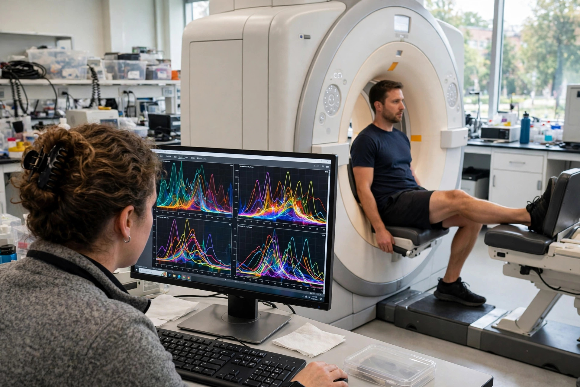
Watching muscle burn fuel in real time: a scanner method tracked mitochondrial pyruvate use during exercise, in 11 men
Almost everything medicine knows about muscle energy comes from biopsies or from measurements taken at rest — while the interesting question is what mitochondria do during work. A study published October 30, 2026 in Nature Communications (Torchi, Mallinson, Serres and colleagues, University of Nottingham) combined two magnetic-resonance methods inside one scanner session: hyperpolarised [1-13C]pyruvate spectroscopy, which follows labelled pyruvate as muscle converts it toward lactate or into mitochondrial metabolism, and phosphorus-31 spectroscopy, which tracks phosphocreatine and pH. Eleven healthy men — four endurance-trained, seven untrained — performed calf (plantar-flexion) exercise inside the scanner at 40% or 70% of their maximum. Harder exercise shifted pyruvate toward lactate relative to mitochondrial use, while trained men showed greater mitochondrial pyruvate flux and less lactate production, mirrored by the phosphorus measures. It is a feasibility study of a measurement method in a small, all-male group — not a clinical test anyone can order, and not a training trial.
Read article →
MitoSwab vs. ME Screen: Which Test Actually Measures Your Mitochondria?
Two at-home mitochondrial tests could hardly measure more different things. MitoSwab assays enzyme activity in your own cheek cells; ME Screen drips your blood serum onto a laboratory cell line and scores how those cells behave. Here is what each test actually measures, what validation stands behind each, and which — if either — is worth $350 or $699.
Read article →
Which aging-clock results actually move? Diet and exercise shifted them across 51 studies; most supplements did not
Epigenetic clocks — blood tests that estimate biological age from DNA methylation — are sold to consumers as report cards for longevity interventions, but until now nobody had run every intervention through the same clocks. The TranslAGE analysis, reported October 29, 2026 from Yale School of Medicine and published in Nature Medicine, did exactly that: 51 human intervention studies, 3,128 samples, each re-scored on the same 16 epigenetic clocks plus 94 methylation measures. Lifestyle interventions combining diet and exercise moved body-aging measures more consistently than most supplements; Mediterranean, low-carbohydrate and low-fat dietary patterns were among those studied. Medicines — metformin, semaglutide and TNF-blocking drugs — also showed effects, largely in patients treated for real disease. Many over-the-counter supplements and procedures showed little measurable effect. The authors' own cautions lead this story: the studies were not head-to-head, and a moved clock is not proven rejuvenation, longer life or fewer illnesses. For mitochondria readers, the pattern is familiar — the interventions that moved the markers are the ones that change cellular energy and inflammation, and the ones that did not are mostly pills marketed as shortcuts around them.
Read article →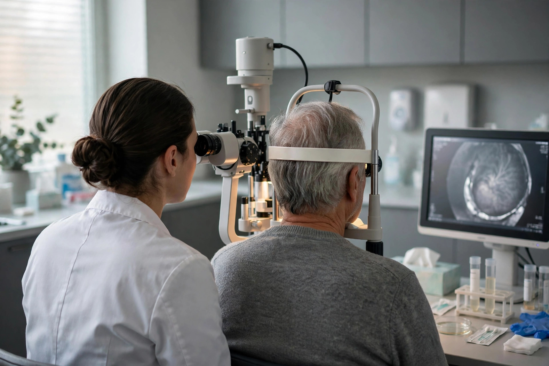
Eye-fluid biopsy maps 64 mitochondrial proteins in geographic atrophy — and an oral nutrient reached the eye
Geographic atrophy — the advanced dry form of age-related macular degeneration — slowly destroys the retina, and researchers cannot biopsy a living retina to ask why. A Stanford-led team (Yeh, Mahajan and colleagues, Communications Medicine, published October 26, 2026) instead sampled aqueous humor, the fluid inside the eye, during routine surgery and profiled it with DNA-aptamer proteomics: 64 mitochondrial proteins were differentially expressed in geographic atrophy, including a coordinated deficiency of the TCA cycle, with reduced PDHB and DLST, two enzymes that control entry into and flux through the cycle. In a separate phase 0 cohort of patients having sequential cataract surgery (ClinicalTrials.gov NCT07269704), one week of oral alpha-ketoglutarate significantly increased intraocular alpha-KG levels and the alpha-KG-to-succinate ratio (P < 0.05), with coordinated shifts across TCA intermediates — the first in-human in vivo evidence that an oral metabolic supplement can engage mitochondrial metabolites inside the eye. No vision benefit was tested or shown.
Read article →
Can a Blood Test Really Grade Your Mitochondria? What the Science Says About Mitochondrial Testing
The short answer: No commercial blood test has been shown to measure your overall "mitochondrial health," and none of the most commonly recommended panels has published the accuracy data a real diagnostic test is expected to have. That is not because the science is uninteresting. It is because "mitochondrial health" has no agreed clinical definition, no reference standard to measure a test against, and — so far — no commercial score that has survived independent checking. What medicine can do well is diagnose mitochondrial disease, and that pathway looks very different from a single number on a lab report.
Read article →
A blood exosome signature classified Parkinson's with high accuracy — and pointed back at mitochondria
Parkinson's still lacks a reliable non-invasive early test. A cross-cohort study in npj Parkinson's Disease, reported October 22, 2026, profiled microRNAs and proteins in blood-derived exosomes — tiny vesicles that can carry material from the brain across the blood-brain barrier. A consensus panel of 69 microRNAs classified Parkinson's strongly in validation, with the best model reaching an AUC of 0.952, while a 63-protein panel reproduced poorly across studies (best AUC 0.611). Across data layers, the biology converged on the same themes: mitochondrial energetics failure and immune dysregulation.
Read article →
Diabetes can hide the mitochondrial signal researchers use to study Alzheimer's
Mitochondrial DNA copy number is a popular stand-in for mitochondrial health in brain research — but it points in opposite directions in two common diseases. A King's College London study in Alzheimer's & Dementia (October 21, 2026) measured mitochondrial DNA copy number in post-mortem hippocampus, amygdala and cerebellum: Alzheimer's was associated with lower levels, diabetes with higher levels. Among people with Alzheimer's, those who also had diabetes had 41% higher levels in the hippocampus, 52% higher in the amygdala and 64% higher in the cerebellum — enough to mask the Alzheimer's signal entirely when samples are pooled.
Read article →
Same mitochondrial gene, many diseases: a 34-patient map shows why the tissue you test matters
MT-CYB is the only complex III subunit encoded by mitochondrial DNA, and its variants can cause anything from exercise intolerance to Leigh syndrome. A Frontiers in Neuroscience study combining a new pediatric case with a systematic review of 34 patients found the variant's share (heteroplasmy) differed sharply by tissue — 22.5% in blood, 70.5% in urine and 95.5% in muscle in the index child — while where variants sat (muscle-restricted vs. widespread) tracked with the clinical picture better than heteroplasmy level itself. For families chasing a diagnosis, a negative blood test does not close the case.
Read article →
What VO2 Max Really Tells You About Your Mitochondria
It is called the gold-standard fitness number, and its final destination is a mitochondrion — every molecule of oxygen you consume ends its journey there. But a VO2 max is a whole-body measurement, and how much of it is really your mitochondria depends on who you are. Here is what the research shows, and how VO2 max stacks up against the tests that measure mitochondria directly.
Read article →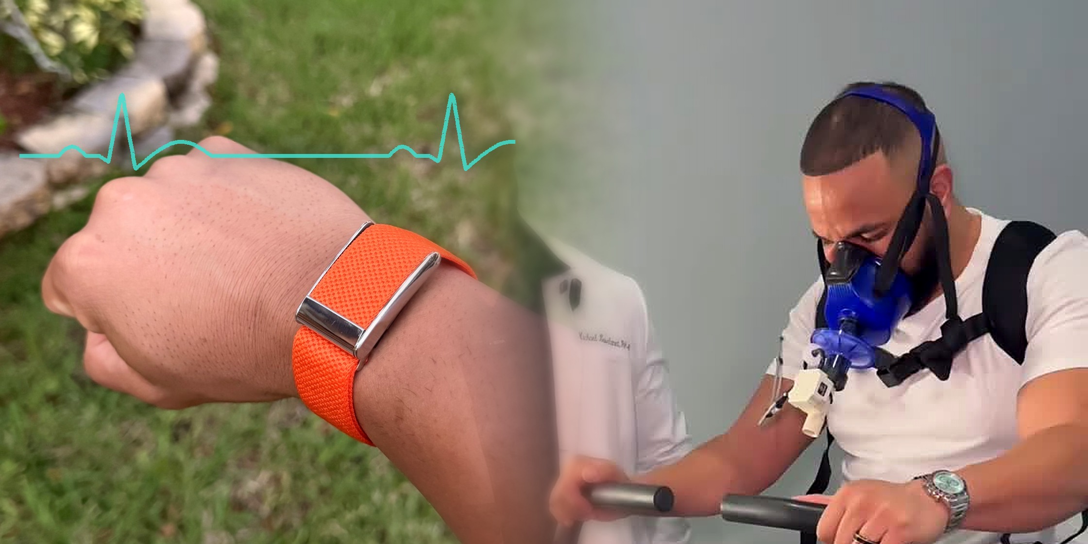
The Estimate and the Measurement: How WHOOP and PNOE Arrive at Your VO2 Max — and Which Number to Trust
Wear a WHOOP band for a few weeks and it will hand you a VO2 max. Book a PNOE test and a technician will hand you one too. The two numbers can differ by five points or more — enough to move you a full fitness category — and the reason is not that one device is broken. It is that they are doing two fundamentally different things. WHOOP estimates your VO2 max from your pulse. PNOE measures it — from your breath and your pulse together, gas exchange and heart rate captured in sync. One is an inference; the other is a measurement. Everything else in this comparison follows from that.
Read article →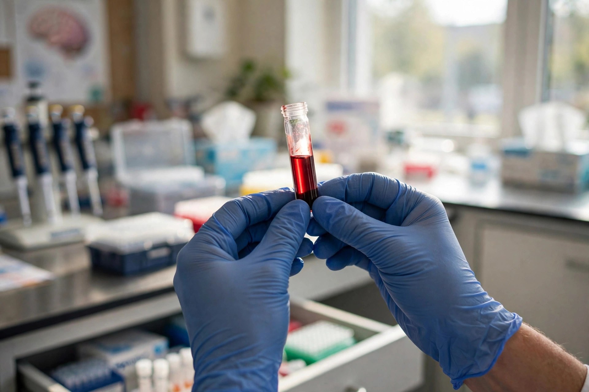
Mitochondrial DNA carried in tiny blood vesicles flagged Alzheimer's memory loss in an early study
Neurons shed microscopic vesicles into the blood, and those vesicles carry mitochondrial DNA from inside the brain. In a Frontiers in Aging Neuroscience study published October 15, levels of that vesicle mtDNA were lower in people with mild cognitive impairment and Alzheimer's — and separated diagnosed Alzheimer's patients from healthy controls with an AUC of 0.965.
Read article →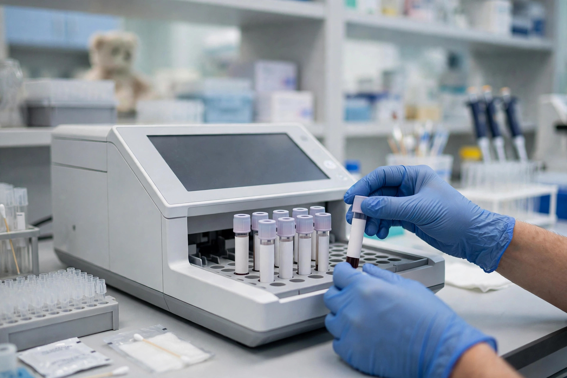
A seven-part blood signature spotted children's mitochondrial disease with high accuracy — in a validation study
Diagnosing mitochondrial disease in children can take years. A study in Nature Communications distilled thousands of blood measurements into a seven-feature panel — six metabolites and one gene transcript — that distinguished affected children from unaffected ones with an AUC of 0.96 in an independent validation group, and a separate twelve-transcript score that predicted how disease would progress.
Read article →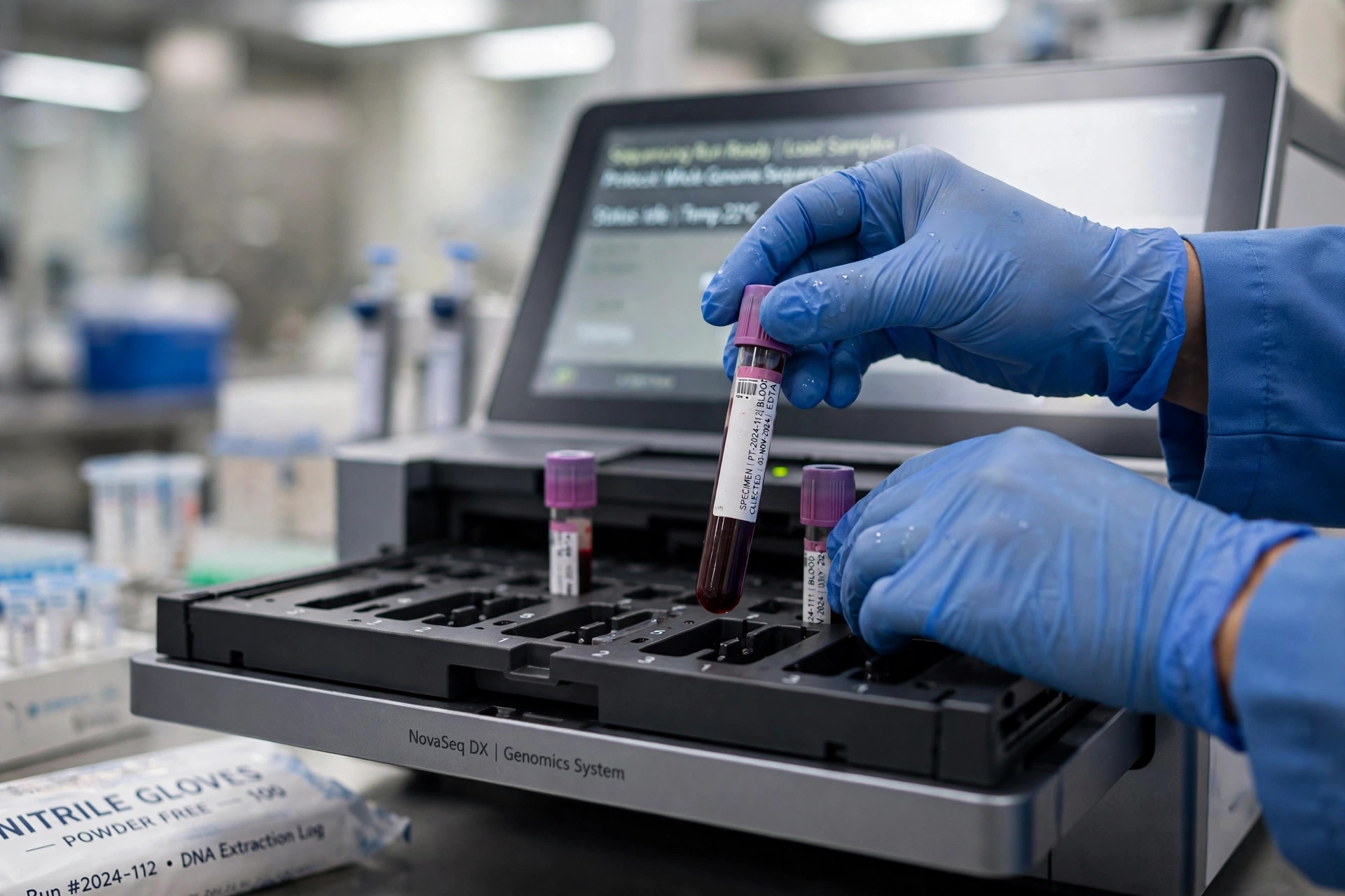
A blood-test fingerprint for schizophrenia and bipolar disorder? Mitochondrial markers show promise
In 934 people from France's I-GIVE cohort, mitochondrial markers (mtDNA copy number, cell-free mtDNA, lactate) plus inflammation signals distinguished schizophrenia and bipolar patients from controls — and moderately separated the two disorders.
Read article →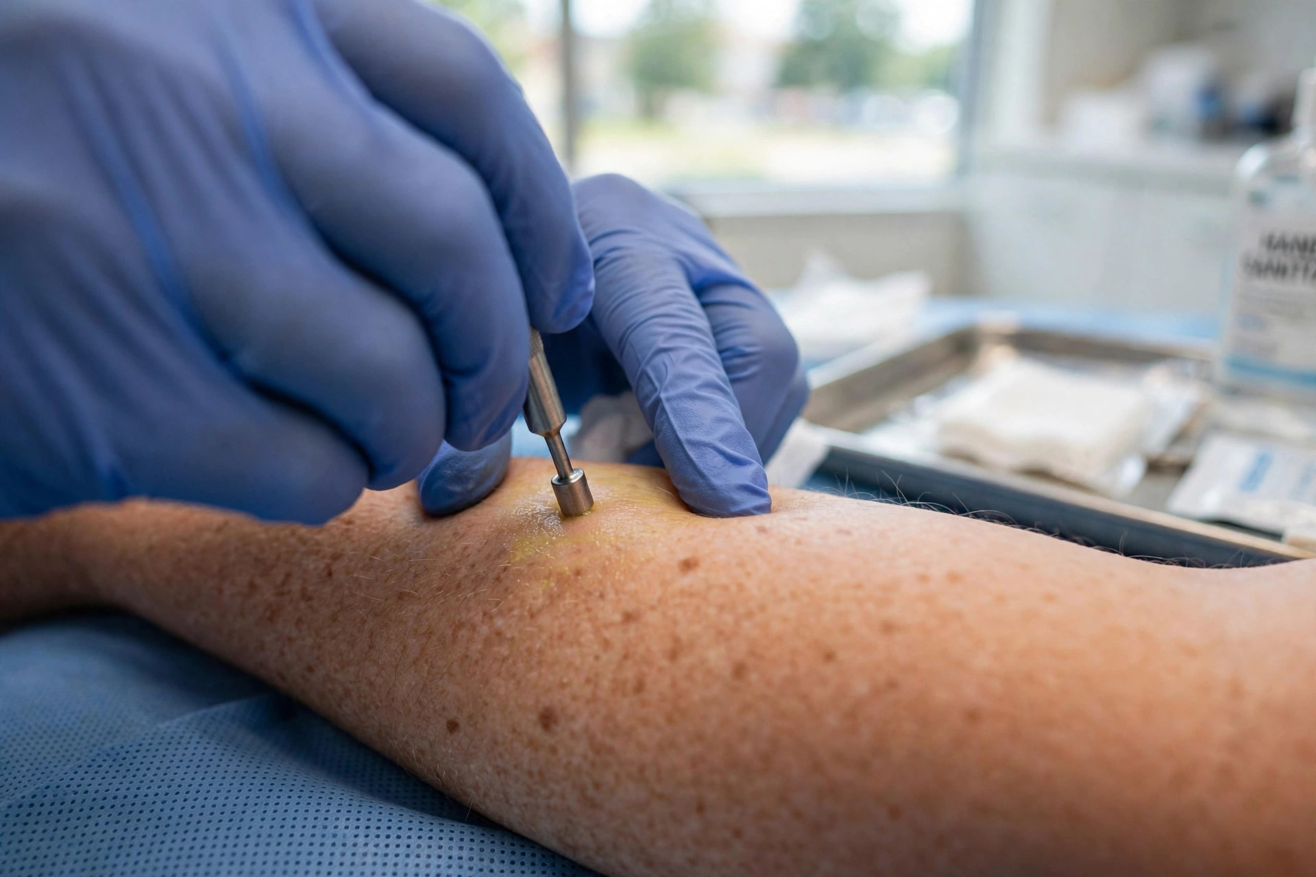
A small skin sample can now help diagnose mitochondrial disease — no muscle biopsy needed
A large international study validates functional testing from skin-biopsy fibroblasts, closing the gap left when genetic tests come back inconclusive — and the tests are now available clinically through Children's Colorado's Mitochondrial Diagnostic Laboratory.
Read article →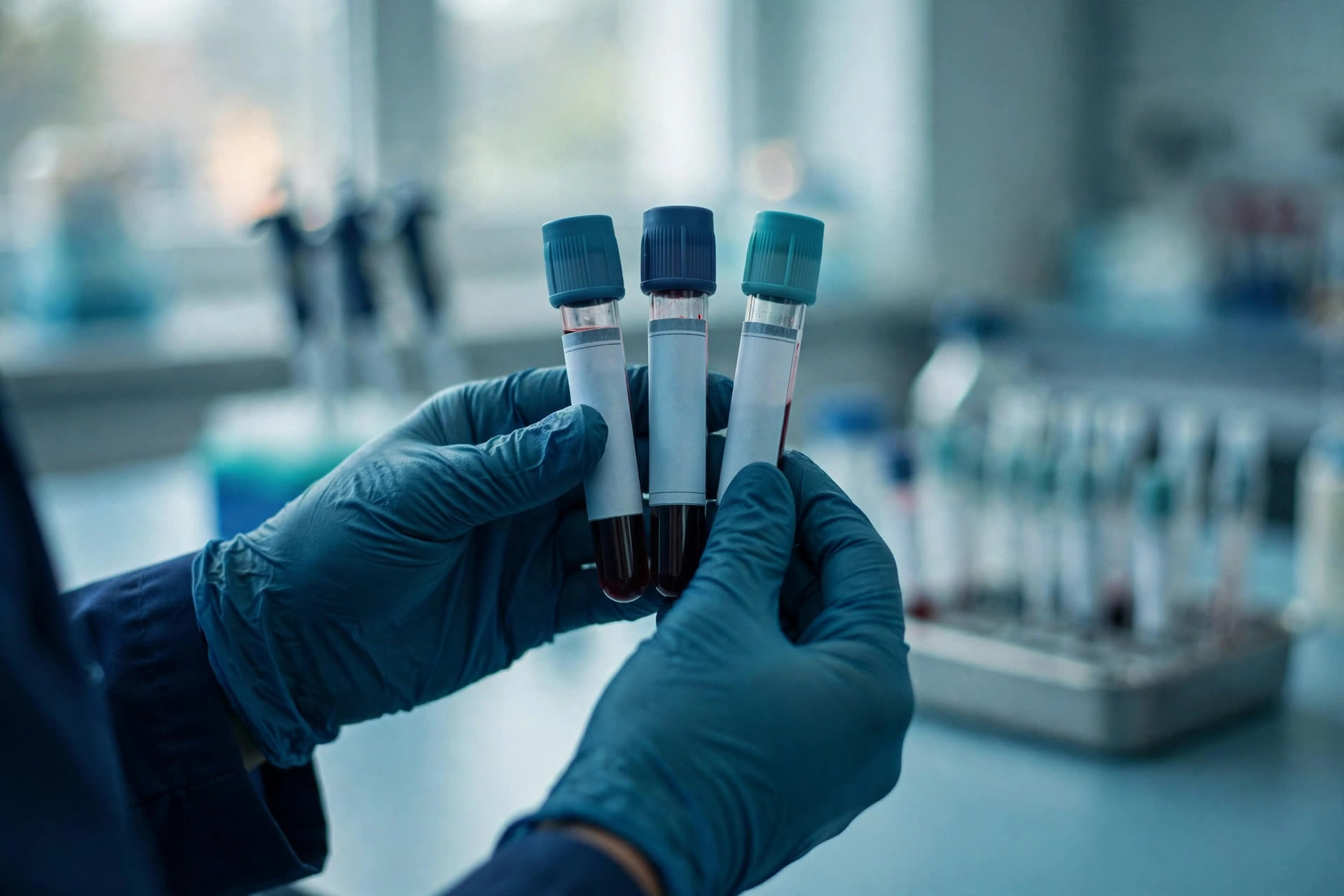
A blood-test clue: mitochondrial DNA copy number separates Parkinson’s from a look-alike disorder
Using quantitative PCR on blood samples, researchers found mitochondrial DNA copy number was significantly lower in Parkinson’s patients than in healthy controls — and also lower than in multiple system atrophy, a neurodegenerative disorder that’s hard to distinguish clinically.
Read article →
Mseek: ultra-pure mitochondrial DNA sequencing
Mount Sinai's Mseek enzymatically deletes nuclear DNA to leave highly pure mtDNA for deep sequencing — a potential step toward using mtDNA as a clinical biomarker.
Read article →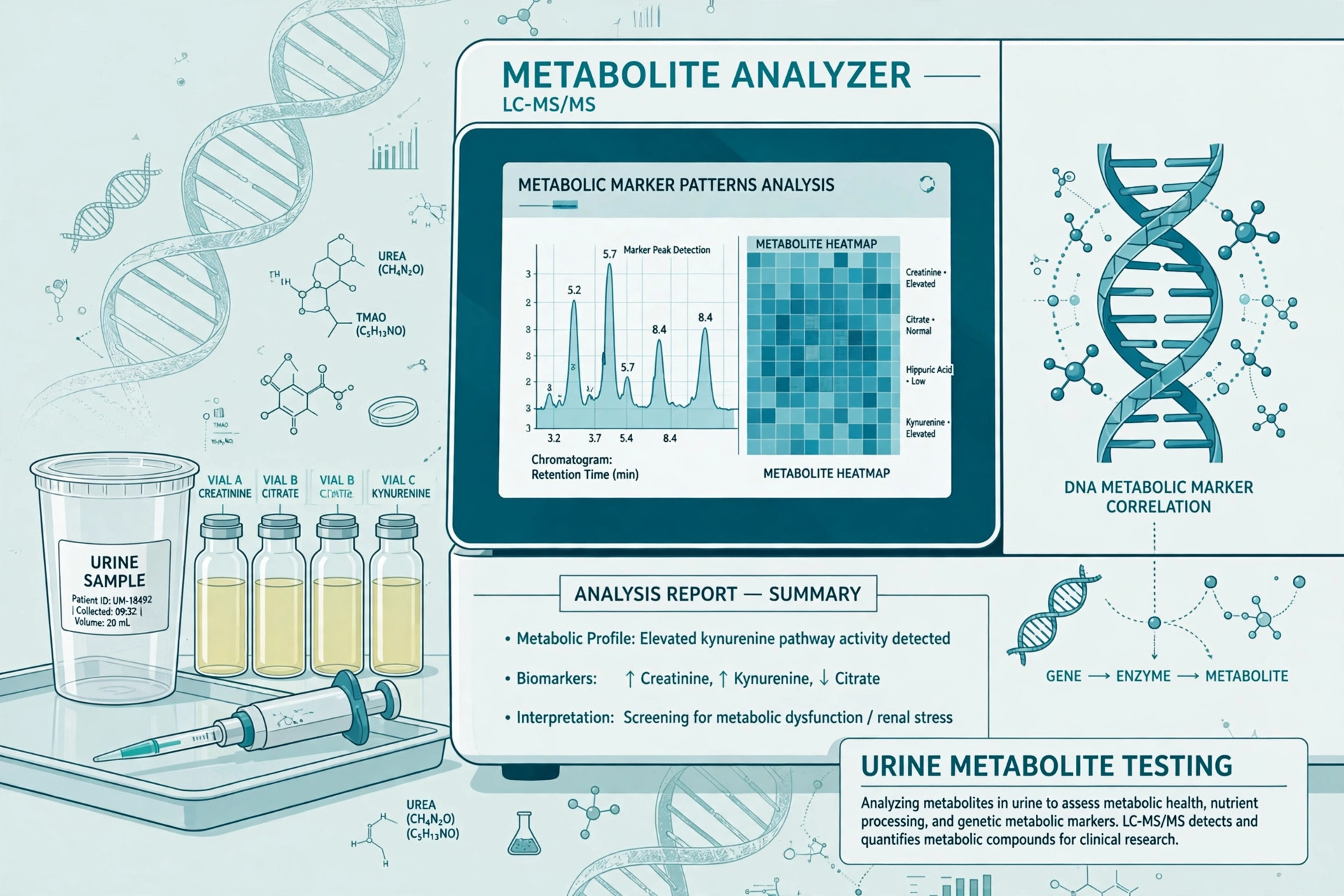
A three-metabolite urine signature may flag a mitochondrial disease variant
A case-control study found a three-metabolite urine signature (uracil, hypoxanthine, 1-methylnicotinamide) distinguishing m.3243A>G carriers with AUC 0.94–0.99. Exploratory; requires validation.
Read article →
What a validated mitochondrial workup measures
A practical guide to mitochondrial DNA sequencing, nuclear gene panels, tissue-level testing, lactate and pyruvate.
Read article →
Inside the IGL mitochondrial test
An evidence review of IGL’s ATP, mitochondrial membrane, antioxidant, nutrient and cellular-toxin panels.
Read article →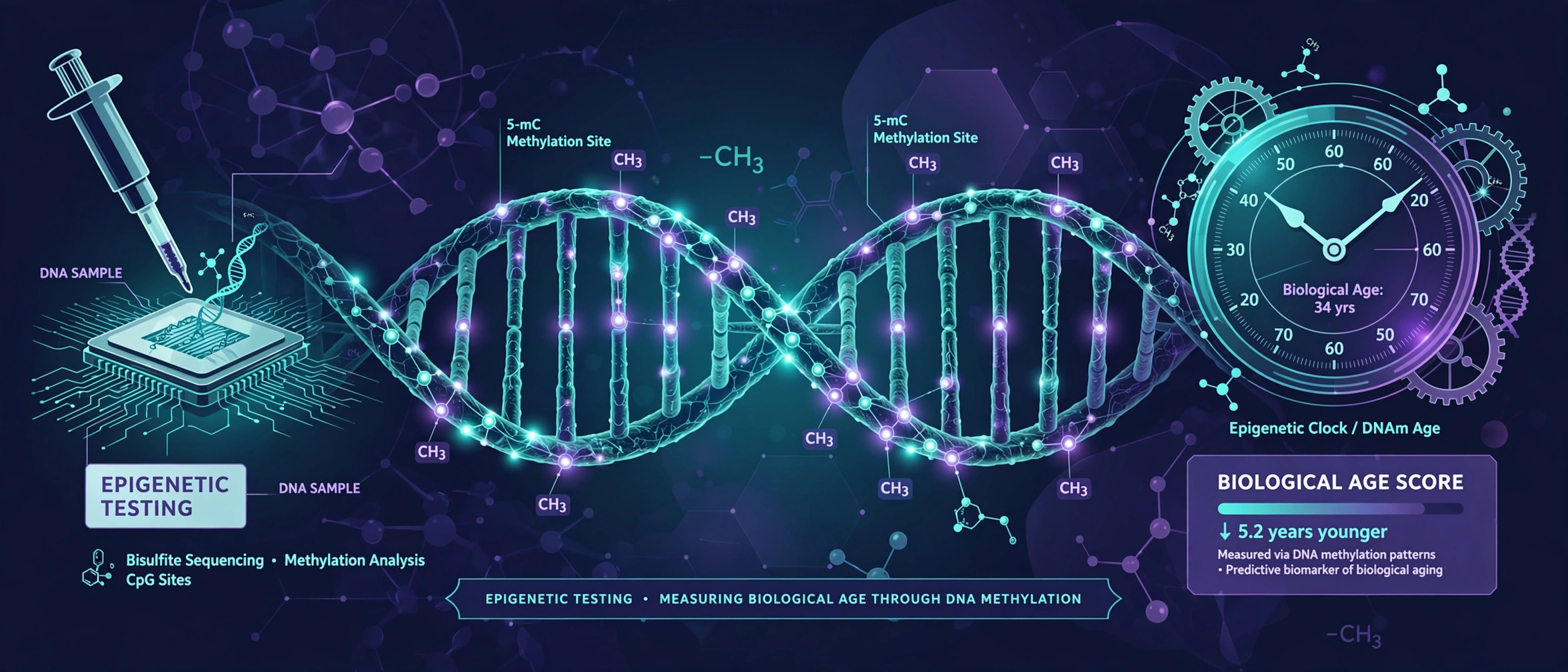
Epigenetic testing and mitochondrial health
What methylation testing can—and cannot—say about mitochondrial genes, function, ATP production and biological aging.
Read article →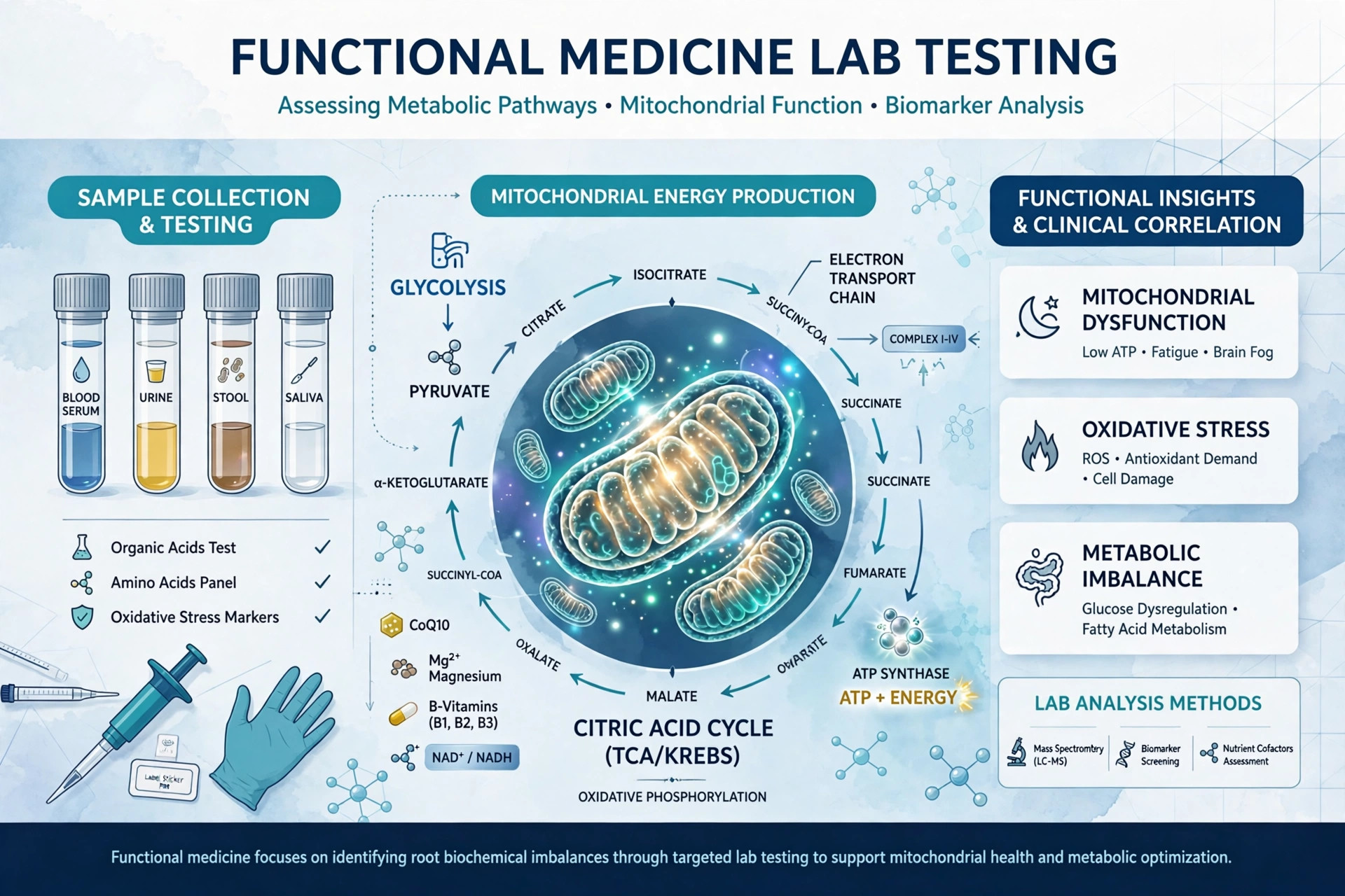
Genova Diagnostics toxin testing
A mitochondria-focused evidence review of Genova toxin, metal, oxidative-stress, genetics and organic-acid panels.
Read article →
Vibrant Wellness Total Tox Burden review
A mitochondria-focused review of Vibrant Wellness urine toxin testing, analytical limits and clinical utility.
Read article →