
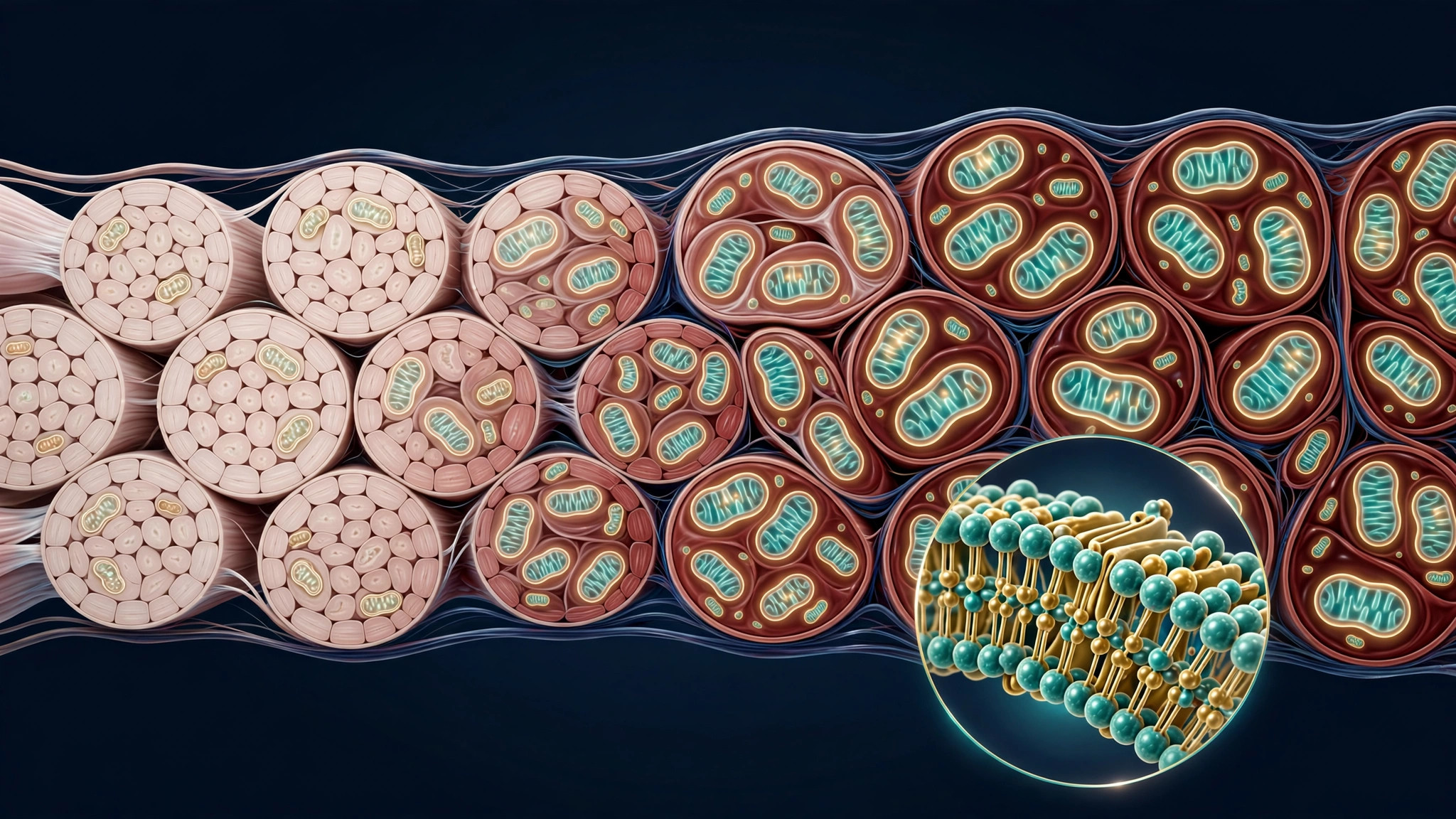
Cardiolipin loss may drive aging-muscle wasting — and restoring it reversed the damage in mice
Aging muscle has a paradox that has puzzled scientists for years: as we age, our muscles shift toward slow-twitch, mitochondria-packed fibers — even as the mitochondria themselves are failing. A new study in Nature Aging, published September 29, finally offers an explanation — and it points to a single fat molecule. Researchers at the University of Copenhagen found that age-related decline in cardiolipin, a lipid found almost exclusively in the inner mitochondrial membrane, drives that fiber shift. And when they partially restored cardiolipin in aged mice, muscle wasting reversed and early deaths were fully prevented.
What is the aging-muscle paradox?
Young muscle is a mix of fast-twitch fibers (powerful, quick to fatigue) and slow-twitch fibers (endurance-oriented, packed with mitochondria). With age, fast-twitch fibers disappear faster — so the remaining muscle looks increasingly slow-twitch and mitochondria-rich. That looks like an adaptation, but muscle keeps wasting anyway. The Copenhagen team — Fabian Finger and Zachary Gerhart-Hines — showed the shift is actually a protective defense response to failing mitochondria, not a sign of failure itself.
How a membrane fat flips the switch
When the researchers lowered cardiolipin in young mice, the fast-to-slow fiber shift appeared on demand. Restoring cardiolipin to roughly two-thirds of normal reversed muscle wasting and fully prevented the early deaths seen in the depleted animals. The switch runs through the nuclear receptor ERRγ: blocking ERRγ in muscle cells shut the fiber shift off completely. Perhaps most surprising, antioxidants made cardiolipin-depleted muscle worse, not better — suggesting the reactive oxygen species are the alarm signal the defense response needs.
Could this become a treatment?
Cardiolipin also declines with age in human muscle samples — but that link is only correlative so far; all the mechanism work was done in mice. Still, the drug angles are real: the FDA recently granted accelerated approval to elamipretide, a cardiolipin-stabilizing peptide, for Barth syndrome, and ERRγ activators are already in preclinical development for other indications. Whether stabilizing cardiolipin helps aging human muscle is an open question — no trial has tested it yet.
Where the evidence stands
- Established: Cardiolipin declines with age in both mouse and human muscle samples, and the fast-to-slow fiber shift with aging is a long-observed phenomenon.
- Preliminary: All causal mechanism work — cardiolipin depletion, partial restoration, ERRγ blockade — was done in mice or muscle cells.
- Contested: Whether restoring cardiolipin helps aging human muscle, and whether elamipretide or ERRγ activators would be safe or effective for that purpose, is not established.
- Absent: No human trial of cardiolipin restoration for age-related muscle wasting exists.