
Cholesterol shuttling keeps lysosomes digesting worn-out mitochondria — in lab models
·Every second, your cells digest their own worn-out mitochondria — a cleanup called mitophagy. The puzzle: how do lysosomes, the acidic recycling centers doing the digesting, survive digesting cargo that big without bursting? A Nature Communications study finds the unlikely answer is cholesterol, rushed in on demand through a molecular supply chain.
What the study found
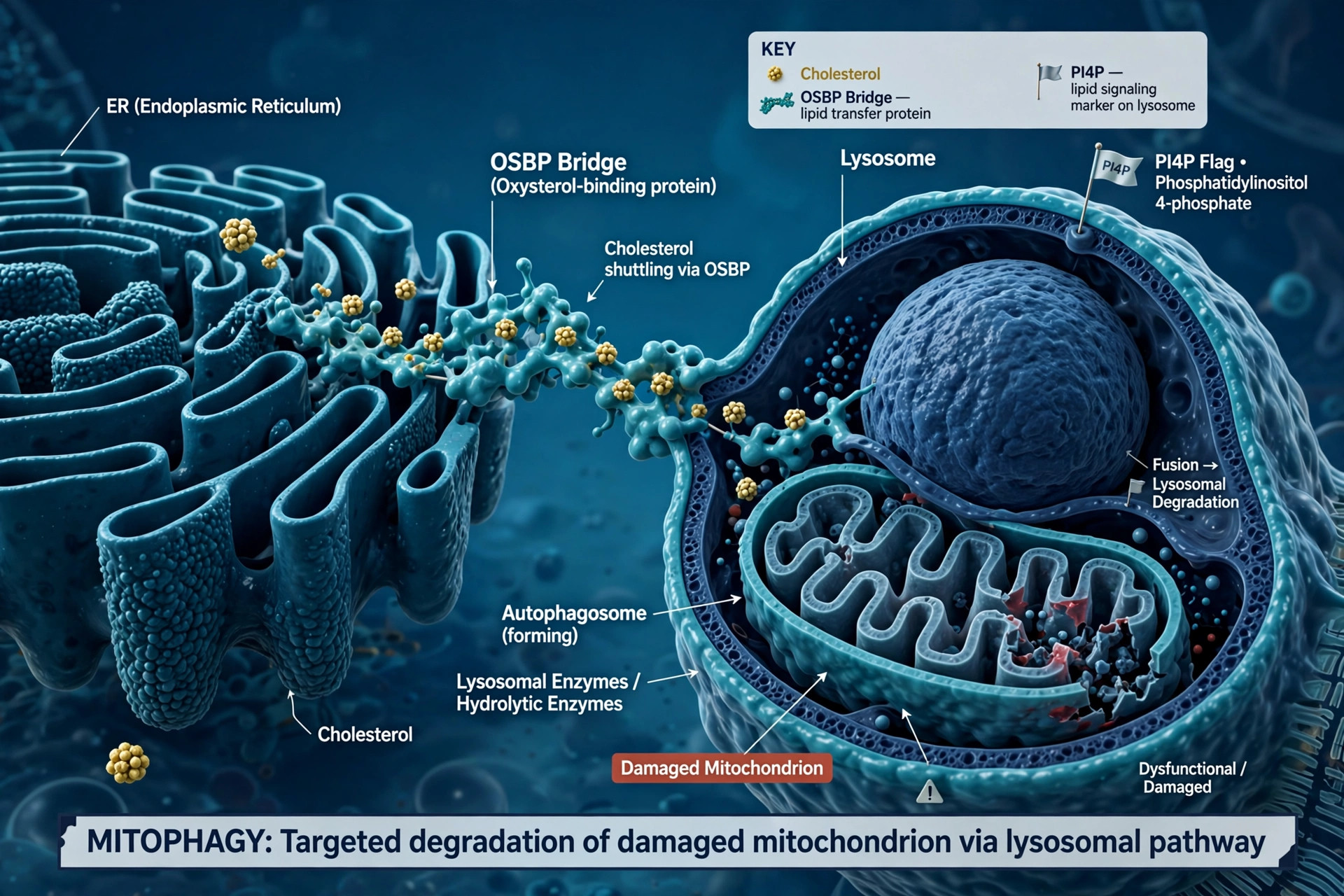
Using live-cell imaging, co-first authors Yang Haoning and Koji Matsuhisa tracked the choreography: a damaged mitochondrion lands in a lysosome; the enzyme PI4KIIα flags the surface with the lipid PI4P; that flag recruits OSBP, a bridge protein that shuttles cholesterol from the endoplasmic reticulum straight into the lysosomal membrane, armoring it against the digestive stress.
The feedback loop
As cholesterol leaves the ER, the cell senses the dip and flips a master genetic switch to make more — keeping the supply chain flowing. Cholesterol thus accumulates precisely where and when mitochondria are depolarized and being degraded.
Why brain aging cares
Failed mitophagy leaves toxic mitochondrial fragments that can trigger cell death — a mechanism implicated in Parkinson's and Alzheimer's. A cholesterol-dependent step in lysosomal resilience is a new place that process can break with age.
The honest limits
This is cell biology in model systems. It identifies a mechanism, not a treatment — and cholesterol handling in the brain is famously double-edged. No intervention follows directly from this paper.
Where the evidence stands
- Established: Mitophagy delivers damaged mitochondria to lysosomes; lysosomal failure contributes to neurodegenerative disease biology.
- Preliminary: The PI4KIIα–PI4P–OSBP cholesterol-shuttle mechanism is demonstrated in cellular models in one study.
- Contested: The net effect of altering cholesterol trafficking in the aging brain — protective versus harmful — is context-dependent and debated.
- Absent: No therapy targets lysosomal cholesterol shuttling for neurodegeneration.