
Scientists found the flexible gate that controls your mitochondria's inner folds
·Tip: press play, then lock your phone or switch apps — the article keeps reading.
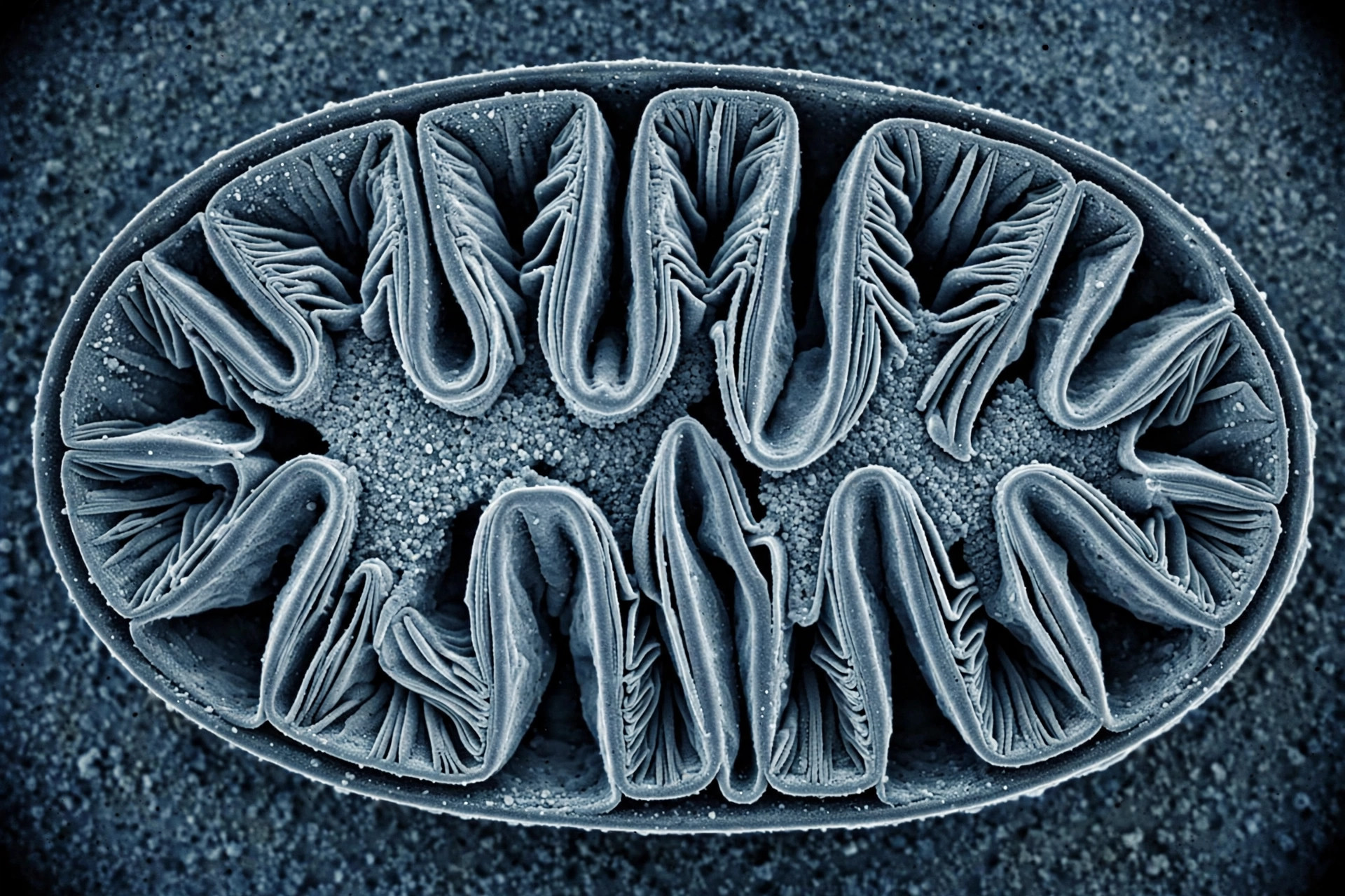
Your mitochondria aren't smooth balloons — their inner membranes fold into elaborate ridges called cristae, and those folds are where energy is actually made. What controls traffic in and out of those folds has been a stubborn mystery. Now, in Nature Communications, researchers have mapped a flexible protein gate — the Mic60-Mic19 subcomplex of the MICOS machinery — that acts as a diffusion barrier at the folds, and shown that a mutation found in human disease alters its core.
What did the researchers find?
Evangelia Nathanail and colleagues used integrative structural modeling to reveal the architecture of the human Mic60-Mic19 subcomplex — the core of the MICOS (mitochondrial contact site and cristae organizing system) machinery. Their model shows this protein pair forming a flexible, dynamic gate that controls which molecules can pass into the cristae folds. Because cristae concentrate the respiratory machinery, anything that governs access to them governs energy production itself.
Computational models are easy to draw and hard to prove, so the team paired the simulations with a new experimental approach for observing the gate in action — a validation strategy they say now gives the field a way to test such models directly.
The disease connection
The finding isn't just structural trivia. The researchers found that a known mutation in patients with optic nerve damage and a developmental brain disorder alters the MICOS complex's core — and their model offers a possible explanation for how that mutation causes disease. Tissues with the highest energy demands, like muscle and brain, are the first to fail when mitochondria falter, which is why a gate defect would strike the optic nerve and the developing brain first.
Confirming the mechanism will require observing the full complex at work inside living mitochondria — so far the team has modeled only part of it — but the study reframes the cristae gate as a genuine therapeutic target rather than an architectural curiosity.
Where the evidence stands
- Established: Cristae folds concentrate the respiratory machinery that produces ATP; high-energy tissues like brain and muscle fail first when mitochondria falter.
- Preliminary: Mic60-Mic19 as a flexible diffusion barrier at cristae — shown by integrative structural modeling plus a new experimental validation method; a disease mutation alters the MICOS core.
- Absent: How the gate behaves in living cells during aging and disease; the full complex observed at work inside mitochondria.