MIC13-linked cristae disruption and mitochondrial liver disease — in lab models
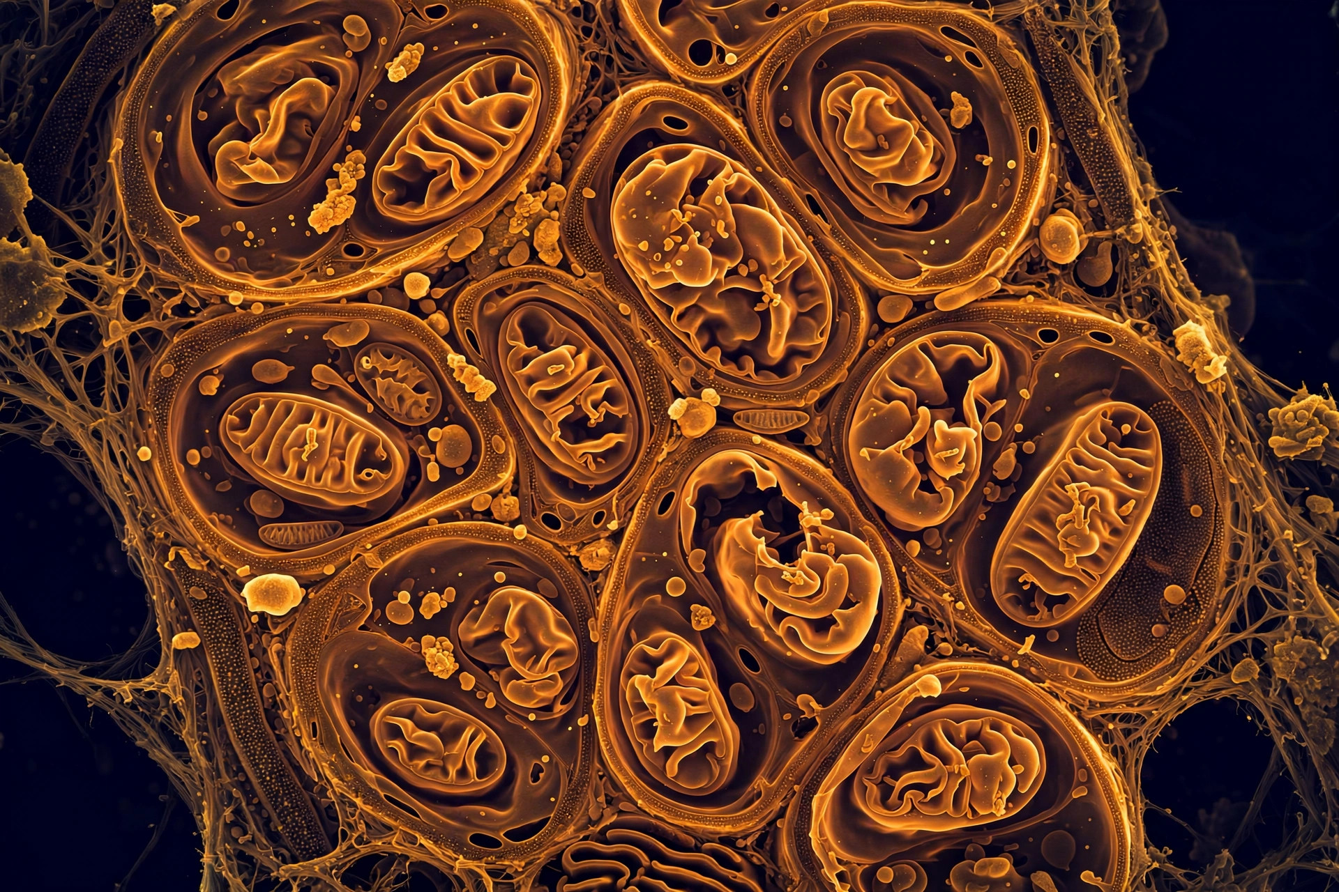
·Your mitochondria's inner membrane is folded into tiny shelves called cristae — and those folds are where the electron transport chain turns food into usable energy. A new study in Cell Death & Disease shows what happens when that architecture collapses: in a cellular model of mitochondrial liver disease, disrupting the cristae-shaping protein MIC13 triggered metabolic failure and early signs of tissue scarring.
Why do cristae matter so much?
Think of cristae as the factory floor of the mitochondrion. The more orderly the folds, the more surface area the respiratory chain has to work with. When MIC13 was disrupted, the folds lost their structure — and energy production paid the price. The researchers documented extensive changes across amino acid, lipid, and energy metabolism.
What went wrong in the model?
The damage did not stay inside the cell. The team found changes in the extracellular matrix — the structural scaffolding around cells — including increased collagen accumulation and altered cell migration. That is the signature of early fibrotic remodeling: the liver beginning to scar. Crucially, this suggests mitochondrial failure can drive tissue-level disease early, rather than merely accompanying advanced damage.
What could this unlock?
The cell model also gives researchers something they have lacked: a workable platform for studying this mitochondriopathy, in a field where research models were previously limited. From here, the hunt is on for the metabolic or molecular vulnerabilities that a future therapy could target. Study DOI: 10.1038/s41419-026-09283-y.
Where the evidence stands
- Established: Cristae architecture is essential for efficient mitochondrial energy production.
- Preliminary: The MIC13-linked mechanism was shown in a cell model; its relevance to human liver disease needs confirmation.
- Absent: No therapy arises from this finding yet.