
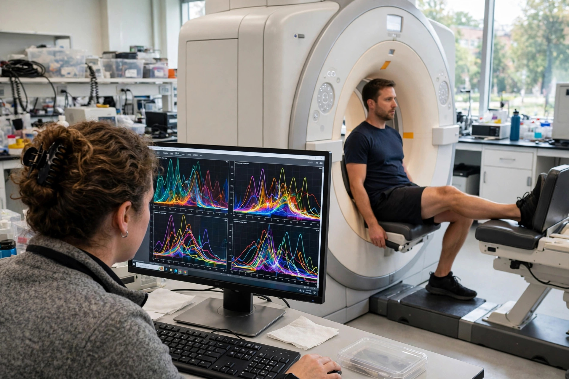
Watching muscle burn fuel in real time: a scanner method tracked mitochondrial pyruvate use during exercise, in 11 men
Edited by Samael A. Tejada, Founder & Editor-in-Chief
·Tip: press play, then lock your phone or switch apps — the article keeps reading.
Researchers combined two scanner methods to watch exercising human muscle metabolism in real time: hyperpolarised carbon-13 pyruvate tracing plus phosphorus-31 spectroscopy. In 11 healthy men, harder exercise shifted pyruvate toward lactate, while endurance-trained men showed greater mitochondrial pyruvate flux. It is a small feasibility study of a research measurement — not a clinical mitochondrial test and not a training trial.
Mitochondrial testing has a standing problem this site returns to often: the claims are about working muscle, but the measurements are usually taken sitting still. This study measured the working muscle itself, while it worked — and showed the trained and untrained patterns differ in the direction physiology predicts. That is what a real measurement advance looks like: narrow, technical, and honest about its size.
How does the method work?
Participants performed steady plantar-flexion exercise inside the scanner. The carbon-13 arm used hyperpolarised [1-13C]pyruvate — pyruvate whose signal is temporarily amplified many-fold so the scanner can follow it — administered intravenously, letting the team watch labelled pyruvate convert to lactate versus entering mitochondrial oxidation in the calf's medial gastrocnemius in real time. The phosphorus-31 arm simultaneously tracked phosphocreatine breakdown and intracellular pH, established measures of muscle energetics. One participant was excluded from the carbon-13 analysis because the tracer was not adequately delivered, leaving ten for that analysis. The magnetic-resonance measurement itself needs no biopsy — though the tracer is given by injection, a distinction this page keeps because 'non-invasive' is often stretched to cover it.
What did trained and untrained muscle do differently?
Two patterns emerged. As exercise intensity rose from 40% to 70% of maximum in untrained men, pyruvate-to-lactate conversion escalated relative to mitochondrial pyruvate utilisation — the expected shift toward faster, oxygen-independent fuel handling under load. Comparing training status at the same relative workload, endurance-trained men showed greater mitochondrial pyruvate flux and reduced lactate production, and the phosphorus measures told the same story from the other side: intensity- and training-dependent differences in phosphocreatine degradation and pH decline. In plain terms, the method could see, live, the metabolic signature that 'better mitochondria' is supposed to mean.
What can readers actually do with this?
Today, nothing directly — and that is the correct answer for a feasibility study. The group was eleven healthy men, the comparison was between people who already differed in training status rather than a randomised training programme, and the platform exists in specialist research centres, not clinics. Its value is as a platform: the authors frame it as a way to phenotype human muscle metabolism during exercise for future studies of ageing, disease and therapeutic interventions — the kind of objective, working-muscle endpoint that mitochondrial drug and exercise trials have lacked. When a commercial 'mitochondrial function test' claims to grade your mitochondria from a resting blood draw, this paper is a useful yardstick for how much machinery a genuine working-muscle measurement currently takes.
Frequently asked questions
What is hyperpolarised pyruvate scanning?
A research MRI method in which pyruvate labelled with carbon-13 is given intravenously with its scanner signal temporarily amplified, so researchers can watch in real time whether muscle turns it into lactate or feeds it into mitochondrial energy production.
What did the study find in trained men?
At the same relative workload, endurance-trained men showed greater mitochondrial pyruvate flux and less lactate production than untrained men, with matching differences in phosphocreatine and pH measured by phosphorus spectroscopy.
Can I get this mitochondrial test at a clinic?
No. It is a specialist research platform demonstrated in 11 healthy men. It is not a validated clinical test, and no commercial service offers it.
Where the evidence stands
- Established: Skeletal muscle energy demand changes rapidly during exercise, limiting the relevance of resting measurements; phosphorus-31 MRS measures of phosphocreatine and pH are established markers of muscle energetics; and endurance training increases muscle mitochondrial oxidative capacity.
- Preliminary: In 11 healthy men (Nature Communications, Oct 30, 2026; DOI 10.1038/s41467-026-79053-z), combined hyperpolarised 13C-pyruvate and 31P spectroscopy during in-scanner exercise showed intensity-dependent shifts toward lactate and, in trained men, greater mitochondrial pyruvate flux with less lactate production.
- Absent: Clinical availability, validation in women, patients or older adults, test-retest standards for individual diagnosis, and any evidence the method changes care or outcomes.
Join the discussion
To get the conversation started: What questions does this story raise for you? Share them below — every comment is reviewed by our moderators before it appears.
Community discussion only — not medical advice. Every comment is reviewed by our moderators before it appears. Sign in with Google or a free Hyvor account to join in.