Two mitochondrial proteins moonlight as disposal tags for damaged mitochondria — and their failure tracks a nerve disease, in cell studies
Edited by Samael A. Tejada, Founder & Editor-in-Chief
·Tip: press play, then lock your phone or switch apps — the article keeps reading.
Quality control is the thread running through modern mitochondrial medicine: cells survive not by having perfect mitochondria but by removing the bad ones quickly. This study names two of the removal tags, shows they sit on proteins already tied to human nerve disease, and demonstrates the disposal failure in patient-derived cells. That is a mechanism with a disease attached — the strongest kind of preclinical story, and still a preclinical one.
What is the second job MFN2 and SLC25A46 do?
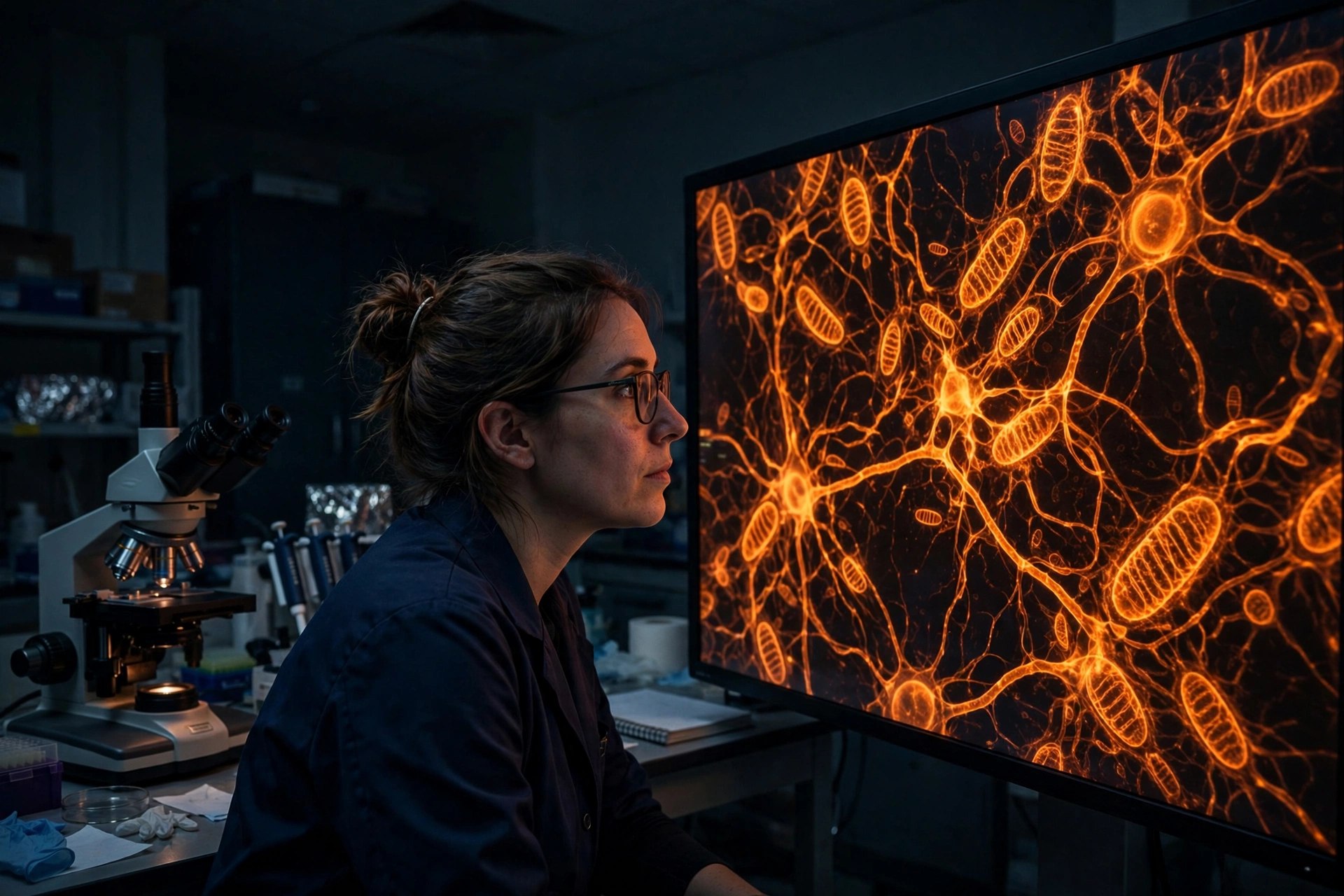
Mitophagy — the selective autophagy of mitochondria — needs receptors: proteins that sit on a damaged mitochondrion and physically recruit the LC3/GABARAP autophagy machinery to engulf it. The Zurich team showed MFN2 and SLC25A46 do exactly that, binding LC3/GABARAP family proteins and serving as receptors on the mitochondrial surface. Both proteins were already known for structural roles — MFN2 drives outer-membrane fusion, SLC25A46 shapes mitochondrial morphology — so the receptor role is an addition to their job descriptions, not a replacement: the same proteins that build the network also help retire its damaged members.
What happened in Charcot-Marie-Tooth patient cells?
Mutations in MFN2 are a known cause of Charcot-Marie-Tooth disease type 2A, an inherited peripheral nerve disease. In fibroblasts taken from patients carrying disease mutations, the team found mitophagy impaired: damaged mitochondria were not cleared at the normal rate and accumulated, and neurons modeled with the defect survived stress less well. That pairing — a clearance mechanism in healthy cells, a clearance failure in patient cells — is what elevates the finding from cell biology to a candidate disease mechanism, and it suggests CMT nerve damage may be partly a waste-disposal disease.
What would a therapy have to prove?
Everything downstream is unbuilt. A drug would need to restore receptor function or bypass it, reach peripheral nerves, clear damaged mitochondria without clearing healthy ones, and show benefit in animals and then patients — none of which exists yet. The near-term value of the paper is diagnostic and strategic: it gives researchers a measurable function (LC3/GABARAP recruitment and mitophagic flux) to screen compounds against, and a reason to test whether other MFN2- and SLC25A46-linked conditions share the same disposal defect.
Frequently asked questions
What is a mitophagy receptor?
A protein on the surface of a damaged mitochondrion that binds the cell's autophagy machinery (LC3/GABARAP proteins) so the organelle is engulfed and recycled. This study shows MFN2 and SLC25A46 perform that role.
How does this connect to Charcot-Marie-Tooth disease?
MFN2 mutations cause Charcot-Marie-Tooth type 2A. In fibroblasts from patients with those mutations, mitophagy was impaired — damaged mitochondria accumulated instead of being cleared — suggesting failed disposal contributes to the nerve disease.
Is there a treatment based on this?
Not yet. The finding is in cells. It identifies a measurable target — restoring mitophagy receptor function — but no drug has been shown to do that in animals or patients.
Where the evidence stands
- Established: Mitophagy clears damaged mitochondria via receptor proteins that recruit LC3/GABARAP autophagy machinery; MFN2 mutations cause Charcot-Marie-Tooth disease type 2A, and impaired mitochondrial quality control is implicated in inherited nerve disease.
- Preliminary: In cell studies (PNAS, Oct 27, 2026; DOI 10.1073/pnas.2521662122), MFN2 and SLC25A46 acted directly as mitophagy receptors binding LC3/GABARAP, and fibroblasts from CMT patients with MFN2 mutations showed impaired clearance of damaged mitochondria and reduced neuronal survival under stress.
- Absent: Animal or human treatment data, any compound that restores the receptor function, and any clinical outcome link in patients.
Join the discussion
To get the conversation started: What questions does this story raise for you? Share them below — every comment is reviewed by our moderators before it appears.
Community discussion only — not medical advice. Every comment is reviewed by our moderators before it appears. Sign in with Google or a free Hyvor account to join in.