
PD-L1, the immunotherapy target, tracks with mitochondrial remodelling in lung cancer cells — in lab-grown cells
Edited by Samael A. Tejada, Founder & Editor-in-Chief
·Tip: press play, then lock your phone or switch apps — the article keeps reading.
In lab-grown non-small cell lung cancer cells, higher PD-L1 expression was associated with altered TCA-cycle metabolic flux, changes in mitochondrial fusion/fission proteins and respiratory-chain components, and distinct epigenetic (H3K9me3) patterns (Cell Death & Disease, November 4, 2026). The study is associational and cell-based: it does not show PD-L1 causes the mitochondrial changes, and no therapy was tested.
PD-L1 is usually photographed at the cell's border, holding up a stop sign to T cells. This study looks inward instead — and finds the cells flying that flag also run differently remodelled mitochondria.
What did the researchers measure?
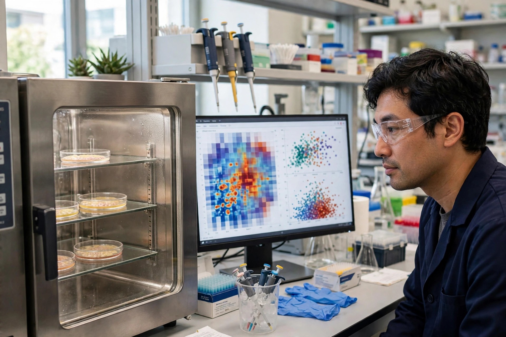
Tumour metabolism and immune evasion are governed by an intertwined network: metabolic dysregulation in tumour cells produces metabolites that can influence immune-cell function, but how PD-L1 expression itself relates to the cancer cell's metabolism has been incompletely understood. Shi and colleagues profiled NSCLC cells differing in PD-L1 expression, combining metabolic flux analysis, mitochondrial protein profiling and epigenomic mapping in an integrated multi-omics design.
What tracked with PD-L1?
Three layers moved together. Metabolically, PD-L1 expression was associated with increased endocytic activity and altered flux particularly through the TCA cycle. Structurally, it was associated with mitochondrial remodelling — alterations in proteins involved in mitochondrial fusion and fission (the constant merging and splitting that shapes the mitochondrial network) and in mitochondrial respiratory-chain components. Epigenetically, multi-omics integration identified differential H3K9me3 patterns between PD-L1 expression states, including at the PTPRN2 locus.
What can't this study tell us?
Direction and consequence. Association in cultured cells cannot say whether PD-L1 drives the metabolic state, the metabolic state drives PD-L1, or both answer to a third signal. It also cannot say whether any of this changes how a patient's cancer responds to PD-L1-blocking immunotherapy — the clinically urgent question this line of work gestures toward. The paper is an early, accepted version subject to further edits, and its conclusions are framed by its own authors as suggestions, not demonstrations.
Frequently asked questions
What is PD-L1?
PD-L1 is a protein some cancers display to suppress immune attack; drugs that block the PD-1/PD-L1 pathway are standard immunotherapies in lung cancer. This study examined what PD-L1 expression tracks with inside the cancer cell's own metabolism.
Did PD-L1 cause the mitochondrial changes?
That is not shown. The study reports associations in cultured lung cancer cells between PD-L1 expression and TCA-cycle flux, mitochondrial fusion/fission proteins, respiratory-chain components and epigenetic patterns. Causation could run in either direction, or both could reflect a shared driver.
Does this change lung cancer treatment?
No. No treatment was tested and no patients were studied. The work is hypothesis-generating for research on why some tumours respond to immunotherapy and others do not.
Where the evidence stands
- Established: PD-L1 is a validated immune-checkpoint target in NSCLC, and tumour metabolic reprogramming — including TCA-cycle rewiring — is established cancer biology.
- Preliminary: The PD-L1–mitochondria association itself: one multi-omics cell study, published as an accepted early version, with no causal or clinical test.
- Absent: Any evidence that targeting mitochondria changes PD-L1 biology or immunotherapy outcomes, and any patient data.
Join the discussion
To get the conversation started: What questions does this story raise for you? Share them below — every comment is reviewed by our moderators before it appears.
Community discussion only — not medical advice. Every comment is reviewed by our moderators before it appears. Sign in with Google or a free Hyvor account to join in.