
Aging rewrites the fat in every tissue differently — but one mitochondrial lipid moves everywhere, in rats
Edited by Samael A. Tejada, Founder & Editor-in-Chief
·Tip: press play, then lock your phone or switch apps — the article keeps reading.
In young versus old male rats, a lipidomics survey of 884 lipids across brain, liver, muscle and plasma found tissue-specific aging patterns, with muscle changing most — and one shared signal: PG, the mitochondrial membrane lipid family, showed the strongest coordinated age-related changes across tissues (Scientific Reports, November 2, 2026). The study is descriptive profiling in rats, with no intervention or human data.
Every tissue ages in its own dialect of fat. This five-tissue census in rats finds the local dialects differ almost completely — except for one mitochondrial lipid family that keeps showing up, in the same direction, nearly everywhere.
What did the five-tissue census find?
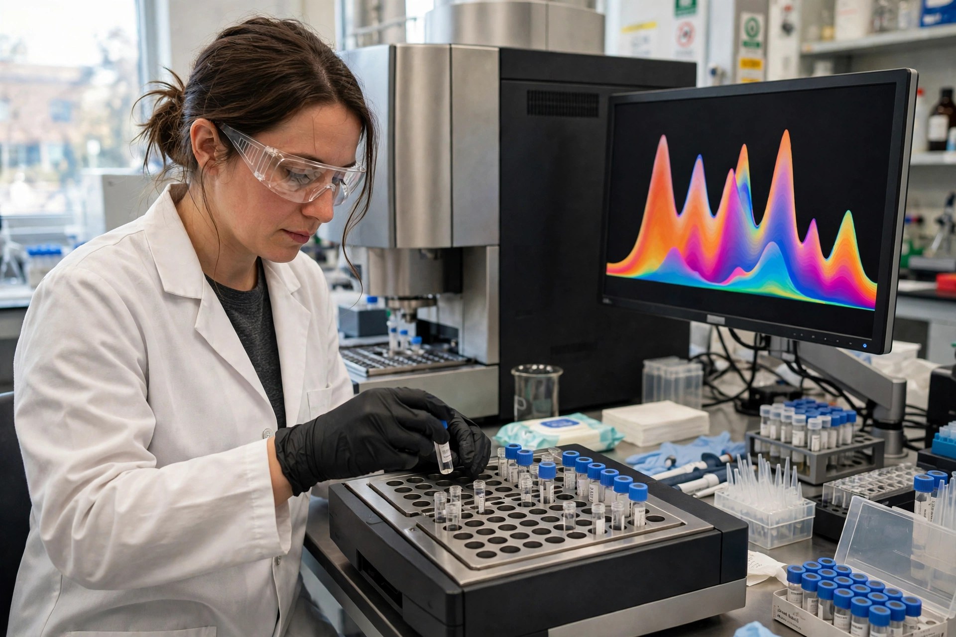
The team profiled frontal cortex, cerebellum, liver, muscle and plasma from young and old male Wistar rats, quantifying 884 lipids in 36 classes. Muscle showed the largest number of changes, with PG and PE O- enrichment the authors interpret as indicating mitochondrial dysfunction. Plasma showed alterations in phosphocholine species (LPC and PC O-), while liver displayed an unexpected decline in triglycerides — particularly shorter- and odd-chain species — consistent with reduced gut microbiota-derived fatty acids. Brain regions showed myelin-related changes: decreased MGDG O- in cerebellum and mixed SHexCer alterations in frontal cortex. Cross-tissue analysis found aging trajectories clustered by tissue function rather than baseline composition, with muscle resembling brain and liver mirroring plasma.
What is the shared mitochondrial signal?
No individual lipid showed ubiquitous age-related change across all tissues — a useful negative result against any single 'aging lipid' biomarker claim. At the class level, though, patterns coordinated, particularly among solid tissues. PG — phosphatidylglycerol, the biosynthetic precursor of cardiolipin, the signature lipid of the inner mitochondrial membrane — showed the strongest coordinated age-related changes. The authors' conclusion is measured: the data support tissue-specific lipid aging processes alongside mitochondrial dysfunction patterns shared across multiple tissues, with mitochondrial lipid remodeling as a common feature of aging. Note what kind of claim that is: coordinated association in a cross-sectional young-versus-old comparison, not a demonstration that changing PG changes aging.
What would make this matter clinically?
Replication in humans, for a start — this cohort is male rats only. Then the harder questions: do the same PG-class shifts appear in human muscle or plasma with age, do they track measured mitochondrial function rather than just birthdays, and does any intervention move them? The plasma findings are the translational hook, since blood is testable in people in a way brain and muscle lipidomes are not. Until those studies exist, this paper's honest contribution is a map: where aging rewrites membrane fat, tissue by tissue, and which mitochondrial corner of that map is shared.
Frequently asked questions
What is PG and why does it point to mitochondria?
PG (phosphatidylglycerol) is a membrane lipid and the precursor family of cardiolipin, the signature lipid of the inner mitochondrial membrane. Coordinated PG changes across tissues are read by the authors as a sign of mitochondrial lipid remodeling with age.
Which tissue changed most with age?
Muscle, in these rats — it showed the largest number of lipidomic changes, with PG and PE O- enrichment indicating mitochondrial dysfunction. Brain, liver and plasma each changed in their own tissue-specific ways.
Is there an aging-lipid blood test from this?
No. Plasma lipids did shift with age in the rats, but no human validation exists and no single lipid changed across all tissues. This is a research map, not a test.
Where the evidence stands
- Established: Mitochondrial membranes are lipid structures whose composition affects function; cardiolipin is the signature inner-mitochondrial-membrane lipid and PG is its precursor family; and aging remodels tissue lipid composition.
- Preliminary: In young vs old male Wistar rats (Scientific Reports, Nov 2, 2026; DOI 10.1038/s41598-026-77040-4), 884 lipids across five compartments showed tissue-specific aging patterns — muscle most changed, with PG/PE O- enrichment — while PG showed the strongest coordinated cross-tissue age-related changes, consistent with shared mitochondrial lipid remodeling.
- Absent: Human data, female animals, longitudinal within-animal tracking, functional mitochondrial measurements tied to the lipid shifts, and any intervention.
Join the discussion
To get the conversation started: What questions does this story raise for you? Share them below — every comment is reviewed by our moderators before it appears.
Community discussion only — not medical advice. Every comment is reviewed by our moderators before it appears. Sign in with Google or a free Hyvor account to join in.