
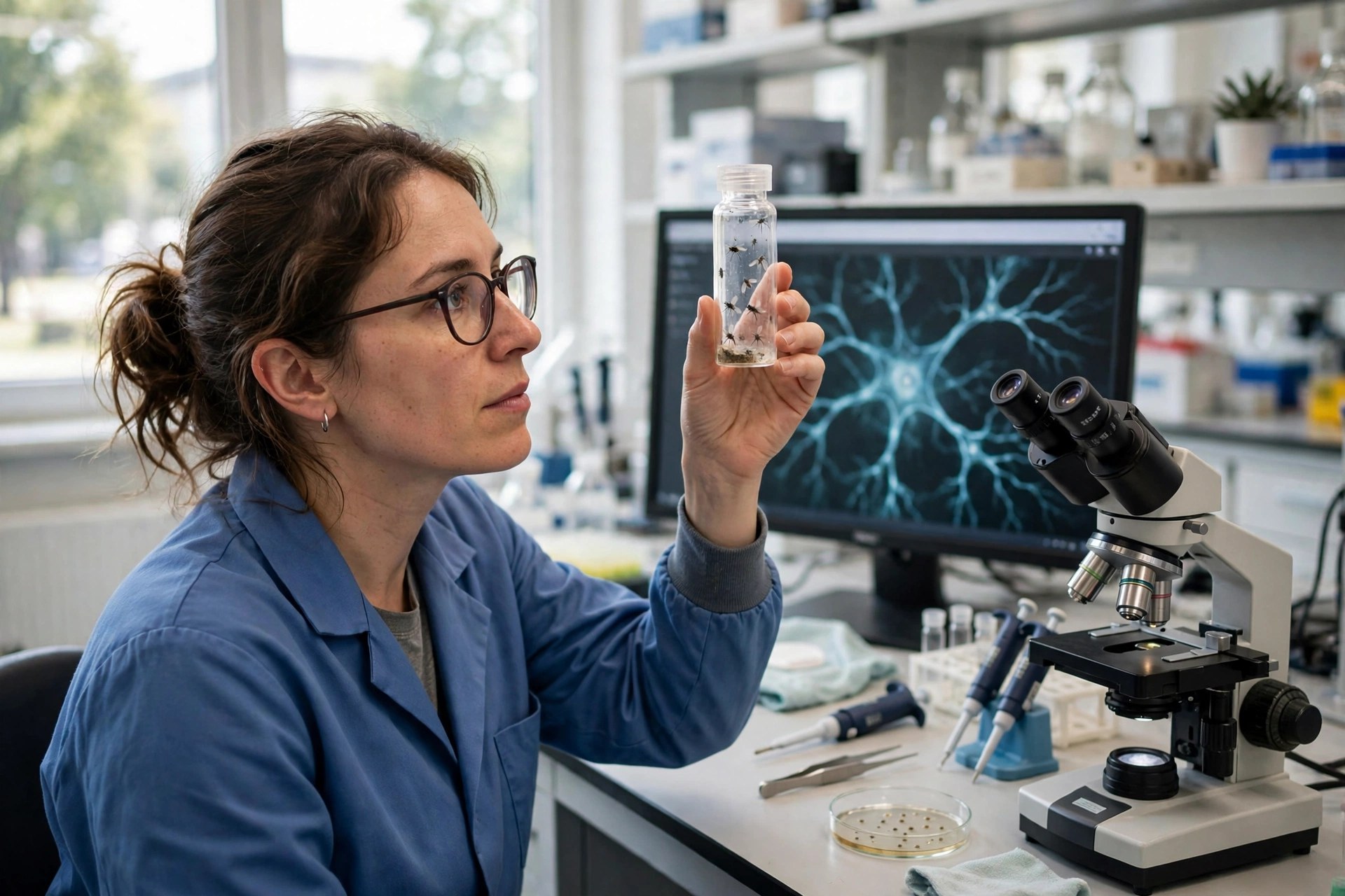
Neurons need their mitochondria to prune their own branches — in fruit flies
Edited by Samael A. Tejada, Founder & Editor-in-Chief
·Tip: press play, then lock your phone or switch apps — the article keeps reading.
In fruit-fly sensory neurons, mitochondria are required for developmental dendrite pruning: disrupting mitochondrial function or transport depleted dendritic mitochondria, prevented caspase activation, and blocked branch severing (Scientific Reports, November 3, 2026). The pro-death proteins Reaper and Grim were also required inside the pruning cell. This is basic developmental biology in flies, with no human or treatment implications tested.
To rebuild itself for adulthood, a fly neuron must first destroy part of itself — precisely, and without dying. This study catches the moment the cutting machinery switches on, and finds the cell's power plants standing in the middle of the operation.
How can a neuron use death enzymes and live?
Caspases are best known as the executioners of apoptosis, but development deploys them sub-lethally for fate specification, migration and differentiation. The fly's dorsal body wall offers a side-by-side comparison in one intact animal: at metamorphosis, class III sensory neurons die by apoptosis while their neighbor, the class IV neuron ddaC, merely prunes its larval dendrites and regrows an adult arbor. With the SR4VH probe, caspase substrates were cleaved in ddaC within 20 minutes of puparium formation — before any branch was severed — at consistently lower levels than in its dying neighbors. Dose, it appears, is the difference between remodeling and death.
What roles do the RHG proteins and mitochondria play?
Single-cell H99 mutant clones showed the proapoptotic RHG proteins are cell-autonomously required for pruning — the signal must come from inside the pruning neuron itself. Although reaper, hid and grim are all transcribed in ddaC as remodeling begins, mutant combinations point to reaper and grim, rather than hid, cooperating to drive branch severing and clearance. Mitochondria sit upstream of the scissors: disrupting mitochondrial function or transport depleted dendritic mitochondria, prevented caspase activation, and blocked both pruning in ddaC and apoptosis in neighboring class III neurons. No mitochondria in the branches, no caspase signal, no cut.
Why does fly pruning matter beyond flies?
Carefully: this is a mechanism paper in an insect model, and it would be wrong to read a human therapy into it. Its value is conceptual and it is real. Developmental pruning is how mammalian brains — including human ones — also refine circuits, and failed or excessive pruning is implicated in neurodevelopmental research. Demonstrating in a living animal that mitochondria gate the caspase signal that severs branches gives that field a concrete, testable mechanism: mitochondrial position and transport inside a dendrite may decide where and whether the cut happens. That is a hypothesis for mammalian work now, not a finding about people.
Frequently asked questions
What is dendrite pruning?
The developmental process by which neurons cut back excess branches so mature circuits wire correctly. In this study, a fruit-fly sensory neuron pruned its larval dendrites at metamorphosis before regrowing an adult-shaped arbor.
What did mitochondria have to do with it?
Disrupting mitochondrial function or transport depleted mitochondria from the dendrites, prevented the caspase activation that drives severing, and blocked pruning — mitochondria are required for the cut, in this fly model.
Does this apply to human brains?
Not directly. Mammalian brains also prune circuits in development, but this study is in fruit flies and tests no human question. It supplies a mechanism for researchers to test in mammalian models.
Where the evidence stands
- Established: Caspases act sub-lethally in development as well as lethally in apoptosis; dendrite pruning is a normal developmental process in Drosophila metamorphosis; and mitochondria are central to caspase activation in cell-death pathways.
- Preliminary: In Drosophila sensory neurons in vivo (Scientific Reports, Nov 3, 2026; DOI 10.1038/s41598-026-77081-9), caspase cleavage began within 20 minutes of puparium formation at sub-lethal levels, Reaper and Grim were cell-autonomously required for pruning, and disrupting mitochondrial function or transport blocked caspase activation, pruning and neighboring apoptosis.
- Absent: Any mammalian or human data, any disease model, and any intervention — this is basic developmental mechanism research in fruit flies.
Join the discussion
To get the conversation started: What questions does this story raise for you? Share them below — every comment is reviewed by our moderators before it appears.
Community discussion only — not medical advice. Every comment is reviewed by our moderators before it appears. Sign in with Google or a free Hyvor account to join in.