
A tumor protein wrecks neutrophils' mitochondria to feed leaky blood vessels in bladder cancer — in lab models
·Tip: press play, then lock your phone or switch apps — the article keeps reading.
Bladder tumors are often flush with blood vessels, yet those vessels are so chaotic they deliver little oxygen, little chemotherapy, and little room for immune cells. A new study in Advanced Science (Chen, Zhang, Xue et al., 2026, vol. 13(56), Article e76604) traces that dysfunction to an unexpected culprit: LRG1 (leucine-rich alpha-2-glycoprotein 1), a secreted tumor protein that hijacks the mitochondria of neutrophils — the short-lived foot soldiers of the immune system — and forces them to release web-like DNA "nets" that corrupt the tumor's blood supply.
What goes wrong with bladder tumor blood vessels?
The team mapped LRG1 across the bladder tumor landscape using four public single-cell RNA sequencing datasets spanning five normal bladder and nineteen bladder cancer samples. One tumor subcluster was defined by high LRG1 output, and its gene program was associated with poorer overall survival in bulk-tumor cohorts from The Cancer Genome Atlas and independent datasets. Patients with blood-borne metastases carried substantially higher LRG1 levels in their serum, and tumor staining showed LRG1 accumulating in the vascular niche of metastatic tumors, where pericyte coverage — the cell wrapping that keeps vessels sealed — was conspicuously reduced. To test causality, the researchers implanted bladder cancer cells into mice genetically lacking Lrg1: tumors grew more slowly, lung metastases were drastically attenuated, and the tumor vasculature underwent vascular normalization — denser pericyte coverage, continuous basement membranes, and wider, calmer vessels.
How does LRG1 break a neutrophil’s mitochondria?
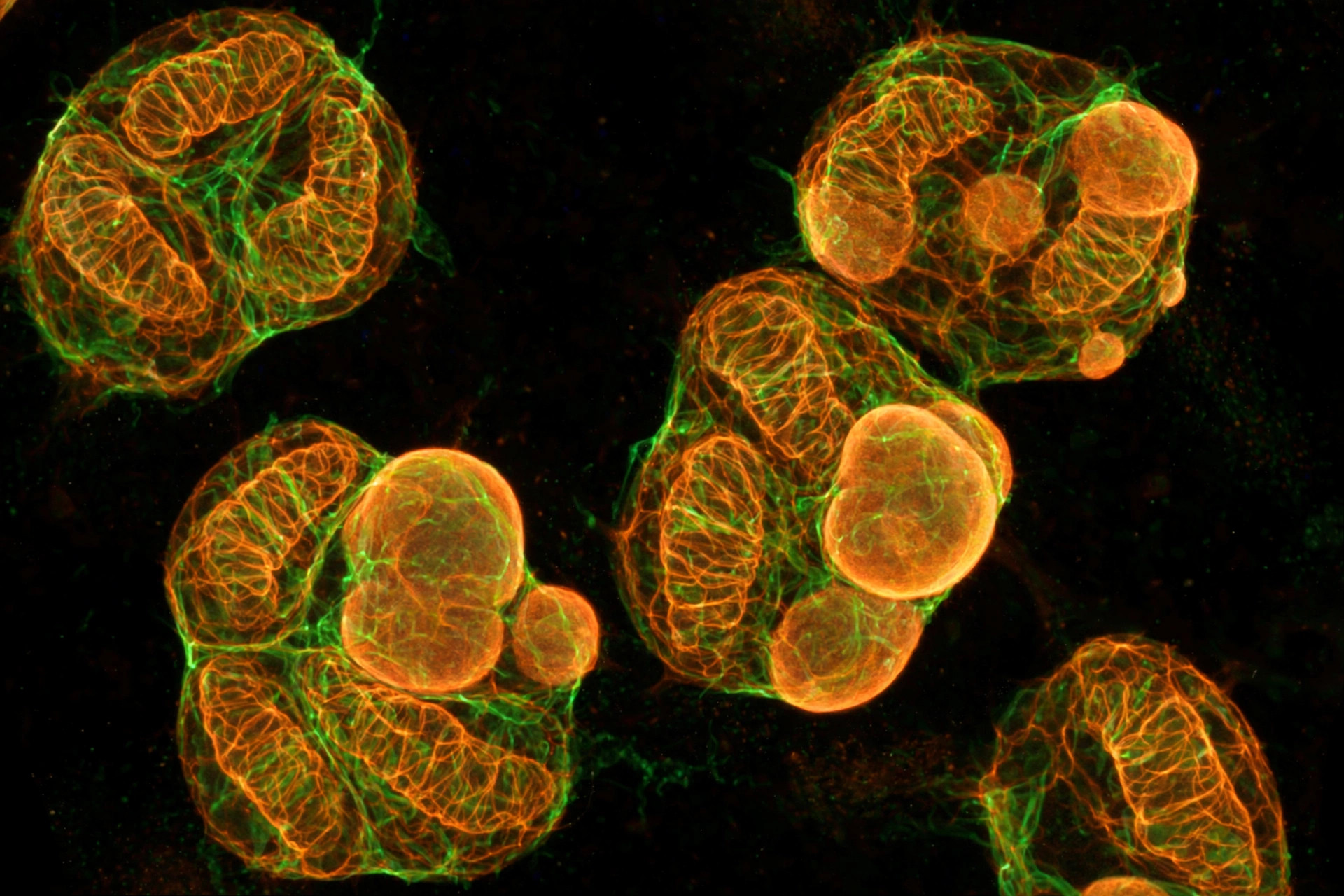
The mitochondrial mechanism is the centerpiece. Using pull-down assays and mass spectrometry, the team identified annexin A2 (ANXA2) as a direct binding partner of LRG1. Normally, ANXA2 ferries activated Akt — a survival signal phosphorylated at serine 473 — into neutrophil mitochondria, a trip that depends on ANXA2's own phosphorylation at tyrosine 24. LRG1 binding blocked ANXA2 phosphorylation and abolished mitochondrial Akt delivery. Starved of that signal, the neutrophils' mitochondria swelled, lost membrane potential, shed mass, spewed reactive oxygen species (mitochondrial ROS — unstable oxygen byproducts that damage what they touch), and failed at ATP synthesis. Transmission electron microscopy showed disorganized cristae — the folded inner-membrane sheets where energy production happens. That mitochondrial collapse pushed neutrophils into NETosis: they exploded outward as neutrophil extracellular traps (NETs), sticky DNA webs loaded with enzymes.
Did blocking LRG1 make treatments work better?
Those NETs attacked the vasculature on two fronts: they accelerated endothelial tube formation while making vessel linings leaky, physically trapped cancer cells, and stripped mural cells from vessel walls. Destroying the NETs' DNA scaffolds with the enzyme DNase I reversed these effects and restored vessel integrity. The translational payoff came in mice: Lrg1 deficiency deepened cisplatin's damage to tumors (better drug penetration) and amplified anti-PD-1 immunotherapy (more CD8 killer T cells with higher Granzyme B). In a retrospective cohort of 29 bladder cancer patients given neoadjuvant cisplatin plus PD-1 blockade, nine of twelve non-responders were LRG1-high and ten of twelve were NET-high. The authors propose the LRG1-ANXA2-NET axis cooperates with the known endothelial TGF-beta/ALK1 pathway to explain bladder cancer's disorganized vessels — and that dismantling it could turn therapy-refractory tumors into ones drugs and immune cells can reach.
The honest catch — and what to watch next
This is strong mechanistic work backed by patient-serum correlations, but the causal experiments are in mice and human neutrophils in dishes. The authors themselves note that neutrophil-specific genetic models are still needed for definitive proof, and the functional role of tumor-specific LRG1 glycosylation is unresolved. LRG1 is also known to act in the eye and other tissues, so any future blocker would need to avoid collateral effects — though the knockout mice showed no major organ abnormalities, an early reassuring sign. There are no clinical trials of LRG1 blockade in bladder cancer yet. Watch for whether human neutrophil mitochondrial ROS — rather than the classical cytosolic route — becomes the confirmed driver of tumor NETosis, and whether LRG1-blocking antibodies already in development for other diseases get tested in this cancer.
Where the evidence stands
- Established: Neutrophils can release DNA-based NETs; bladder cancer is known for leaky, dysfunctional tumor vasculature; mitochondrial ROS can drive NETosis.
- Preliminary: The LRG1-ANXA2-mitochondrial Akt axis, mitochondrial damage in neutrophils, vascular normalization from LRG1 loss, and improved chemo/immunotherapy responses were shown in mouse bladder-cancer models and human neutrophils in vitro. Patient-serum LRG1 correlations and a 29-patient retrospective cohort are associative, not causal.
- Absent: Any human clinical trial of LRG1 blockade in bladder cancer, neutrophil-specific genetic confirmation of the NETosis link, and safety data for targeting LRG1 systemically.
Join the discussion
To get the conversation started: What questions does this story raise for you? Share them below — every comment is reviewed by our moderators before it appears.
Community discussion only — not medical advice. Every comment is reviewed by our moderators before it appears. Sign in with Google or a free Hyvor account to join in.