
A clearer way to watch mitochondria inside hard-to-see nerve-support cells — in mice
·Tip: press play, then lock your phone or switch apps — the article keeps reading.
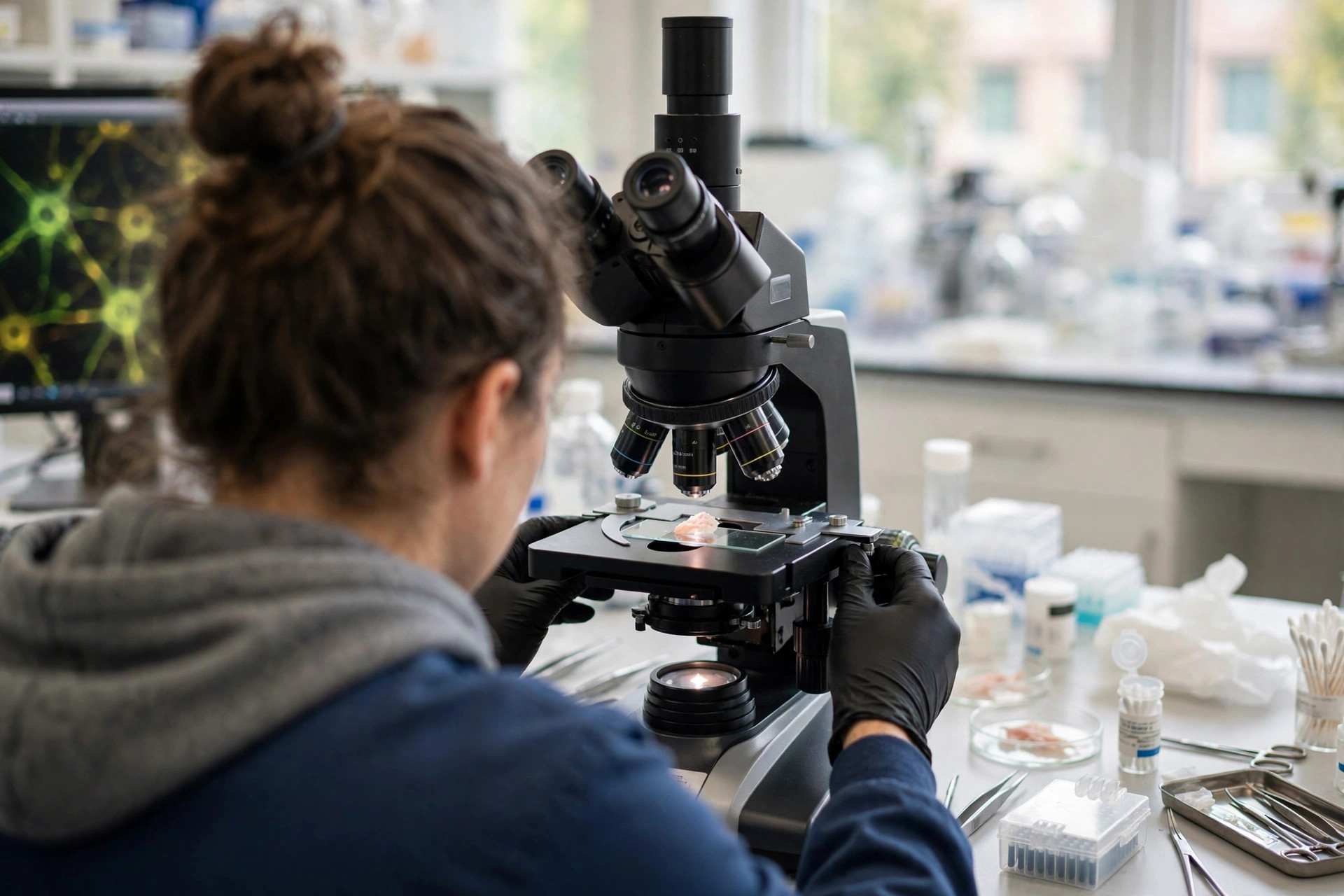
Every voluntary movement ends at the neuromuscular junction, where a motor nerve meets muscle. Capping that junction sit terminal Schwann cells, support cells implicated in ALS, diabetic neuropathy, and spinal muscular atrophy — yet historically understudied, largely because researchers lacked good ways to examine them in their natural setting. A methods study published in the Journal of Visualized Experiments (Morton and colleagues) sets out to close that tooling gap for one specific question: what do these cells' mitochondria look like, and how are they organized?
What does the method do?
The protocol lets researchers quantify mitochondrial morphology — size, shape, and network organization — in situ, inside intact mouse muscle and terminal Schwann cells, rather than in isolated cells grown in a dish. Because mitochondrial shape tracks closely with mitochondrial health and function, being able to measure it in the cells' real environment gives a more faithful picture of how these support cells are doing.
Does it actually detect meaningful differences?
In the team's demonstration, yes: the technique captured differences in mitochondrial organization between healthy and dystrophic terminal Schwann cells — in the researchers' plain description, healthy support cells keep their mitochondria networked together, while stressed ones fragment and keep to themselves. Showing that a measurement can separate a known healthy state from a known disease state is the basic test a new method has to pass before it can be trusted on unknown questions.
Why publish a tool as news?
Because in this corner of neuroscience, the tool has been the bottleneck. With a validated way to read mitochondrial organization in terminal Schwann cells, researchers can now ask disease questions that were previously impractical — including, in this lab's own plans, whether an experimental biomaterial designed to promote mitochondrial health improves nerve recovery after injury. No disease was treated and no patient is affected today; the contribution is a sharper instrument for the studies that come next.
Where the evidence stands
- Established: Terminal Schwann cells support the neuromuscular junction and have been implicated in ALS, diabetic neuropathy, and spinal muscular atrophy; mitochondrial shape is a recognized readout of mitochondrial health.
- Preliminary: A new in-situ protocol quantified mitochondrial morphology in mouse muscle and terminal Schwann cells and detected organization differences between healthy and dystrophic tissue.
- Absent: Application of the method to human tissue, any disease-mechanism finding produced with it so far, and results from the planned mitochondrial-biomaterial nerve-recovery experiments.
Join the discussion
To get the conversation started: What questions does this story raise for you? Share them below — every comment is reviewed by our moderators before it appears.
Community discussion only — not medical advice. Every comment is reviewed by our moderators before it appears. Sign in with Google or a free Hyvor account to join in.