A new microscope watches mitochondria change shape and chemistry at the same time — demonstrated in lab-grown cancer cells
Edited by Samael A. Tejada, Founder & Editor-in-Chief
·Tip: press play, then lock your phone or switch apps — the article keeps reading.
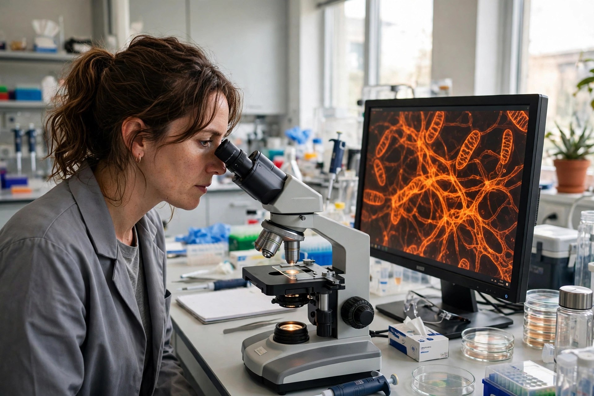
A new microscopy method, SIMFLIM, combines super-resolution structured illumination microscopy with fluorescence lifetime imaging to show both the fine structure of organelles like mitochondria and their molecular environment (such as pH-sensitive changes) simultaneously, at high speed (Optica, 2026; announced November 5, 2026). It was demonstrated in a human bone cancer cell line; it is a research tool, not a clinical test.
A microscope that only shows shape can miss the chemistry, and one that only shows chemistry can miss the shape. For mitochondria — organelles that constantly remodel while their internal chemistry shifts — that split view has been a real blind spot.
How does SIMFLIM work?
Structured illumination microscopy beats the normal resolution limit of light microscopy, making visible cellular structures too small to resolve conventionally. Fluorescence lifetime imaging measures a different quantity: how long fluorescent molecules emit light after excitation, which changes depending on their molecular environment — for example, due to changes in pH. Until now, combining the two at high resolution has been possible only to a limited extent. The new FLIM-SIM combination adds high spatial resolution, fast imaging rates and a large field of view, and the team demonstrated it on a human bone cancer cell line; the published images include mitochondria changing structure over time.
Why does drug research want this?
The CHEM-SCAN project is investigating how chemotherapeutic agents can be adapted to act as effectively as possible on both tumours and their metastases. Specialized fluorescent dyes, read through FLIM measurements, report on the state of cancer cells. 'By combining super-resolution with fluorescence lifetime measurements, we can not only see where certain molecules are located within a cell. We also gain information about the molecular environment in which they are located,' says Dr. Henning Ortkraß, one of the study's authors, in the group's announcement. Project coordinator Professor Thomas Huser adds that the results 'open up a new dimension of contrast for super-resolution microscopy,' with live-cell and drug research the intended applications.
Is this a mitochondrial test patients will encounter?
No. SIMFLIM is a laboratory research method for studying cells — including how their mitochondria behave under drugs — not a clinical test of anyone's mitochondrial health. Its relevance to readers is upstream: better measurement tools are how mitochondrial drug effects get verified or debunked long before anything reaches a clinic, and methods papers like this one set the standard of evidence the field can demand.
Frequently asked questions
What is SIMFLIM?
SIMFLIM is the combination of structured illumination microscopy (SIM), a super-resolution imaging technique, with fluorescence lifetime imaging microscopy (FLIM), which measures how long fluorescent molecules glow and thereby reports on their molecular environment.
What does it show about mitochondria?
In the demonstration images, it resolves mitochondrial structures changing over time while simultaneously providing fluorescence-lifetime contrast — information about the molecular surroundings, such as pH-related changes — that structure alone cannot give.
Can this test my mitochondrial health?
No. It is a research microscope method for studying cells in laboratories, developed under the European CHEM-SCAN project for drug research. It is not a clinical or consumer test.
Where the evidence stands
- Established: SIM and FLIM are each established microscopy techniques; fluorescence lifetime genuinely reports on molecular environment such as pH.
- Preliminary: The combination's performance: one methods paper (Optica, 2026) with demonstrations in a human bone cancer cell line; adoption and benchmarking by other laboratories will decide its standing.
- Absent: Any clinical application. This is not a diagnostic or consumer mitochondrial test.
Join the discussion
To get the conversation started: What questions does this story raise for you? Share them below — every comment is reviewed by our moderators before it appears.
Community discussion only — not medical advice. Every comment is reviewed by our moderators before it appears. Sign in with Google or a free Hyvor account to join in.