
What VO2 Max Really Tells You About Your Mitochondria
·Tip: press play, then lock your phone or switch apps — the article keeps reading.
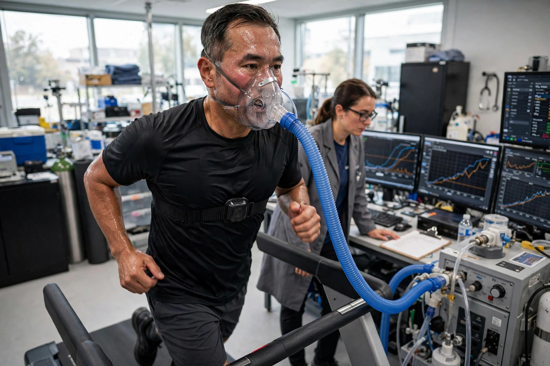
Ask around the longevity world and you will hear it stated as fact: VO2 max is one of the best diagnostic tools we have for mitochondrial health. The claim has a real foundation. VO2 max — the maximum rate at which your body can use oxygen during all-out exercise, measured with a mask and a metabolic cart — is the strongest single predictor of future health and survival ever validated in exercise science. And because oxygen's last stop is the mitochondrion, the number is genuinely connected to your cellular engines.
The claim, and the short answer
But precision matters here, because two different claims are being fused into one. VO2 max is an outstanding *integrative* test: it grades the entire oxygen delivery-and-use system, and it predicts outcomes better than almost anything else in medicine. It is not, however, a *mitochondrial* test in the way a biopsy is. It cannot isolate your mitochondria, it cannot locate a defect, and in the fittest people it may say surprisingly little about them. Understanding where the connection holds — and where it breaks — is the difference between using the number well and being misled by it.
Why your VO2 max ends at a mitochondrion
Follow a breath of oxygen. It enters the lungs, crosses into the blood, binds to haemoglobin, and is pumped by the heart through arteries to working muscle. There it leaves the capillaries, crosses into the muscle cell, and arrives at the mitochondrion — specifically at complex IV of the electron transport chain, the last step of the machinery that turns food and oxygen into ATP, your cells' usable energy. Oxygen is consumed nowhere else in the body in any meaningful amount. Mitochondria are the sink at the end of the pipeline.
Physiologists express the whole system in one line, the Fick equation: VO2 max equals maximal cardiac output multiplied by the maximal difference in oxygen content between arterial and venous blood — how much the heart delivers, times how much the tissues extract. The American Heart Association's scientific statement on fitness describes the same chain: lungs, heart, blood vessels and muscle cells each contribute. The mitochondrial implication is exact but subtle: at maximal exercise, your body can never consume oxygen faster than your muscle mitochondria can burn it. Mitochondria set the ceiling of demand. Whether your measured VO2 max actually reaches that ceiling depends on whether the delivery side of the pipeline can keep up — and that is where the story gets interesting.
Where the link is strong
When researchers compare VO2 max against direct measures of muscle mitochondria, the relationship is real:
- Breathing muscle fibres in a chamber. In a 2002 *Journal of Physiology* study, researchers biopsied thigh muscle from sedentary, active and athletic adults and measured how fast the fibres consumed oxygen. The fibres' maximal respiration tracked whole-body peak VO2 with a correlation of r = 0.63 — and rose in a clean staircase with fitness: sedentary muscle burned 3.7 units of oxygen, active muscle 5.9, athletic muscle 7.9.
- Across the full human range. A 2016 study spanning chronic heart failure patients to professional cyclists — VO2 max values from 9.8 to 79.0 ml/kg/min — found that mitochondrial oxidative capacity estimated from muscle staining predicted measured VO2 max with an r-squared of 0.89, and that people on average used about 90 percent of their predicted mitochondrial capacity.
- In the untrained. In a 2016 study of young men, the mitochondrial capacity of biopsied muscle matched and predicted whole-body VO2 max almost perfectly in untrained men (r = 0.91). Their mitochondria genuinely were the limiting factor.
The pattern underneath all three: when the range of fitness is wide, or when people are sedentary, VO2 max and muscle mitochondrial capacity move together. In that setting, a low VO2 max really does carry information about the mitochondria in your muscles.
The catch: a fit heart hides its mitochondria
The same 2016 study shows where the link breaks. In endurance-trained men, the mitochondria in a muscle biopsy could consume oxygen at a rate of 743 ml/kg/min — but the body in action reached only 458. The trained mitochondria had roughly 60 percent reserve capacity that the heart and blood vessels simply could not feed. In trained people, the bottleneck is delivery — cardiac output, blood volume, haemoglobin — not the mitochondria. The classic review of this literature (Bassett and Howley, 2000) makes the point several ways: raise or lower oxygen delivery — with blood doping, altitude or beta-blockers — and VO2 max moves with it, no mitochondrial change required. Double a person's mitochondrial enzymes with training and VO2 max rises only 20 to 40 percent. Two athletes with identical VO2 max scores can differ two-fold in mitochondrial enzyme levels.
The dissociations run in both directions. A 2021 *Journal of Physiology* study found that VO2 peak falls with age even though the maximal respiration of biopsied muscle fibres did not differ between young and older adults — the age-related fitness decline in that cohort was a delivery story, not a mitochondrial one. And a meta-analysis of transfusion and blood-donation studies found VO2 max tracks haemoglobin concentration almost perfectly (R-squared = 0.99): change the blood, change the number, mitochondria untouched.
None of this makes VO2 max a bad test. It makes it a *system* test. What it grades is the whole pipeline — and the mitochondria are one section of that pipeline, sometimes the limiting one, sometimes not.
The number that predicts how long you live
Whatever VO2 max is measuring, it predicts the future with a strength few biomarkers in medicine can match:
- Per unit of fitness, risk falls steadily. A 2009 meta-analysis in *JAMA* covering 102,980 people found that each 1-MET increase in aerobic capacity — a modest, achievable fitness gain — was associated with 13 percent lower all-cause mortality and 15 percent fewer coronary and cardiovascular events.
- The fittest live dramatically longer. In a 2018 *JAMA Network Open* study of 122,007 treadmill-tested adults followed for a median of 8.4 years, elite fitness was associated with an adjusted mortality hazard of 0.20 versus the lowest fitness group — an 80 percent lower risk of death — with no upper limit of benefit observed, even at the highest fitness levels.
- Medicine has noticed. The American Heart Association's 2016 scientific statement argues cardiorespiratory fitness should be assessed as a clinical *vital sign*, noting low fitness rivals or exceeds smoking, hypertension, high cholesterol and type 2 diabetes as a risk factor. A 2024 overview of meta-analyses pooling 20.9 million observations confirmed the pattern: per 1-MET higher fitness, 11 to 17 percent lower mortality and 18 percent lower risk of heart failure.
One caution of interpretation: none of that prognostic power proves the signal is mitochondrial. VO2 max predicts outcomes precisely *because* it integrates heart, lungs, blood, muscle, body composition and habitual activity into one number. Borrowing those mortality statistics to sell VO2 max as a mitochondrial diagnostic mixes up two different kinds of validity — predicting the future, versus measuring a specific organelle.
Does it move when mitochondria improve?
Broadly, yes — in parallel, but loosely. Across 97 training groups in a systematic review, the relative rise in citrate synthase — a standard marker of mitochondrial content in muscle — correlated with the relative rise in VO2 max at r = 0.45: real, but explaining only about a fifth of the variation. Training studies repeatedly show mitochondrial content climbing far faster than VO2 max — content up 40 to 55 percent in six weeks in one biopsy study while VO2 max rose far less — because much of the VO2 max gain comes from the heart and blood, not the muscle.
The most telling data come from people whose mitochondria are the problem. In patients with mitochondrial myopathy, endurance training raised VO2 max by roughly a quarter across studies — and in a 2006 *Brain* study the 11 percent VO2 gain came with an 11 percent gain in oxygen *extraction*, the muscle-side term. In these patients, unlike healthy trainees, a rising VO2 max really is a mitochondrial story.
When a low VO2 max really is a mitochondrial signal
In primary mitochondrial disease — genetic defects of the respiratory chain — exercise testing earns a genuine diagnostic role, though a supporting one. Patients typically show low peak VO2, an exaggerated heart-rate and breathing response for the workload, and crucially, a failure to extract oxygen: the blood leaving exercising muscle stays unusually rich in oxygen because the mitochondria cannot use what arrives. The degree of impairment tracks the proportion of mutant mitochondrial DNA in muscle. Exercise protocols separate mitochondrial myopathy patients from controls with sensitivity around 0.63 to 0.75 and specificity around 0.70 to 0.90 — useful, not decisive. More specific extraction-based indices from the CPET report perform better in small studies, but none is prospectively validated at scale, and confirmation always comes from genetics and tissue testing.
Contrast that with heart failure, the other great clinical use of VO2 peak: there the same low number (below 10 to 14 ml/kg/min marks the highest-risk Weber classes) usually means the *heart* cannot deliver — the opposite mechanism. Same test, same low number, opposite implication for the mitochondria. That ambiguity is the honest summary of VO2 max as a diagnostic: powerful, and never self-interpreting.
The tests that measure mitochondria directly
How does the exercise test compare with the tools that aim at the mitochondria themselves?
Muscle biopsy with respirometry — the reference standard. A small sample of thigh muscle is biopsied, its fibres permeabilised, and their oxygen consumption measured chamber by chamber (high-resolution respirometry, e.g. the Oroboros Oxygraph), alongside enzyme assays for citrate synthase and each respiratory complex. Citrate synthase activity matches the electron-microscope gold standard for mitochondrial content at r = 0.84. Nothing else reads muscle mitochondrial capacity this directly. The costs: it is invasive, samples one small site, measures capacity under laboratory conditions rather than life, and lives in research and specialist centres — there is no routine clinical price.
Phosphorus-31 MRS — the in-vivo direct measure. Inside a specialised MRI scanner, researchers watch phosphocreatine — the muscle's rapid-energy buffer — rebuild after exercise. Because that rebuilding is purely oxidative, its time constant (about 25 to 35 seconds in healthy calf muscle) indexes mitochondrial capacity in living muscle, and it validates against biopsy measures. But it needs scarce multinuclear scanner time and expertise, and for *diagnosing* mitochondrial myopathy it performs surprisingly poorly (specificity below 65 percent in the 2021 review) — an excellent physiological measure can still be a weak diagnostic test.
NIRS — the portable proxy. Near-infrared light through the skin tracks muscle oxygen use; with brief cuff occlusions after exercise, its recovery rate constant matches the 31P-MRS number closely (r = 0.88 to 0.95, identical average time constants of 31.5 seconds in the cross-validation study). It is non-invasive, portable and far cheaper — but confounded by fat thickness and blood volume, standardised for research rather than clinic, and it measures one small muscle region.
Lactate — cheap, everywhere, non-specific. Lactate rises when glycolysis outruns mitochondrial oxidation, so it whispers about mitochondria without ever naming them. In suspected mitochondrial disease, resting lactate is only 34 to 62 percent sensitive (though 83 to 100 percent specific), and a supervised lactate stress test improves that to about 0.67 sensitivity and 0.93 specificity in a 2024 meta-analysis. Many things raise lactate — including a tight tourniquet and a clenched fist during the blood draw — and many confirmed mitochondrial patients test normal. Best used as context inside an exercise test, never as a standalone verdict.
GDF15 and FGF21 — the blood stress signals. These "mitokines" are released when mitochondria activate the cellular stress response, and for diagnosing primary mitochondrial *disease* they post the best blood numbers available: a 2020 meta-analysis found GDF15 sensitivity 0.83, specificity 0.92; FGF21 sensitivity 0.71, specificity 0.88. But both rise with age, cancer, cardiovascular and kidney disease, and metformin; published cut-offs vary nearly ten-fold; and neither tells a healthy person anything quantitative about their respiratory capacity. Supportive evidence in a disease work-up — not a mitochondrial health score.
Blood-cell respirometry — the cautionary tale. The appealing idea: measure respiration in blood cells (PBMCs or platelets, on a Seahorse analyser) as a liquid biopsy of your mitochondria. The largest check, a 2024 study with 318 blood samples and paired muscle biopsies, found neither blood cell type's respiration correlated significantly with muscle respiration — and athletes' superior muscle mitochondria were not mirrored in their blood. Gross genetic defects can show up in blood; ordinary variation does not. Research only.
Skin fibroblast testing — the validator. A small skin punch, cultured for weeks, lets specialist labs run the full functional panel — enzyme activities, complex assembly, protein abundance. A large multicentre study reported in 2026 found the combined panel 76 percent sensitive and 93 percent specific for primary mitochondrial disease — though it caught only 20 percent of one disease category (mtDNA-maintenance disorders), and it is a single recent study awaiting replication. Remember its name: this is the Children's Colorado work in our earlier coverage of skin-biopsy diagnosis. Its modern role is resolving uncertain genetic findings, not screening the healthy.
Genetic testing — the cause, not the capacity. Mitochondrial and nuclear gene panels answer a question no functional test can: *is there an inherited defect, and which one?* That guides prognosis, family risk and drug safety. But DNA says nothing about how your mitochondria are performing today — blood can miss tissue-specific mutation loads, mutation percentage in blood poorly predicts severity, and acquired dysfunction from inactivity, illness or medication leaves no trace in the sequence.
The sensible way to use it
Put together, expert guidance converges on a stack. Start with the exercise test: a measured VO2 max (see our companion piece on WHOOP versus PNOE for how the number is obtained, and why measurement beats estimation) gives you the best-validated picture of your integrated aerobic system and your risk trajectory, for roughly $199 to $600 at US clinics by current listings. Track it over time, same protocol, same device — trend is where its mitochondrial information lives, because in sedentary and deconditioned people, gains in VO2 max and gains in muscle mitochondria genuinely travel together. If a local muscle number matters — in research or training monitoring — NIRS recovery is the evidence-backed non-invasive option. And if the question is diagnostic — symptoms, family history, suspected disease — VO2 max becomes one supporting witness among several: mitokine blood markers, genetics first for cause, and biopsy or fibroblast functional testing when tissue answers will change care.
What VO2 max cannot do, no matter how it is marketed: give a healthy person a mitochondrial diagnosis, grade their mitochondria on a scale, or substitute for the tests that measure the organelle itself. There is no validated VO2 max cut-off for mitochondrial dysfunction in healthy people, and no blood test today returns a trustworthy mitochondrial function score. The number deserves its reputation — as the best whole-body window we have on the system your mitochondria anchor, and arguably the most meaningful single number in preventive medicine. Just read it for what it is.
Where the evidence stands
- Established: VO2 max / cardiorespiratory fitness predicts mortality and cardiovascular events with unusual strength (per 1-MET: roughly 13% lower all-cause mortality; elite vs lowest fitness HR 0.20) — the basis of the AHA "fitness as a vital sign" statement.
- Established: In sedentary and untrained people, and across wide fitness ranges, VO2 max correlates substantially with direct measures of muscle mitochondrial capacity (r = 0.63 to 0.91 in biopsy studies).
- Established: In trained people VO2 max is limited mainly by oxygen delivery; mitochondrial capacity exceeds what the circulation can supply, so VO2 max under-reports mitochondrial capacity.
- Established: Muscle biopsy respirometry and 31P-MRS phosphocreatine recovery measure mitochondrial function directly; GDF15/FGF21 are validated supportive biomarkers for primary mitochondrial disease.
- Preliminary: NIRS muscle oxygen recovery as a clinical tool (validated against 31P-MRS in research; no clinical cut-offs); fibroblast panel performance (one large 2026 study); CPET extraction indices for mitochondrial myopathy (small single-centre studies).
- Contested: Whether ageing itself lowers intrinsic muscle mitochondrial respiration, or mainly reflects delivery and activity changes (direct dissociation data exist); whether type 2 diabetes impairs mitochondrial quality versus quantity.
- Absent: Any validated VO2 max cut-off for diagnosing mitochondrial dysfunction in healthy people; any blood test (including PBMC/platelet respirometry, cell-free mtDNA, metabolomics) validated as a mitochondrial function score for healthy people.
Join the discussion
To get the conversation started: What questions does this story raise for you? Share them below — every comment is reviewed by our moderators before it appears.
Community discussion only — not medical advice. Every comment is reviewed by our moderators before it appears. Sign in with Google or a free Hyvor account to join in.