
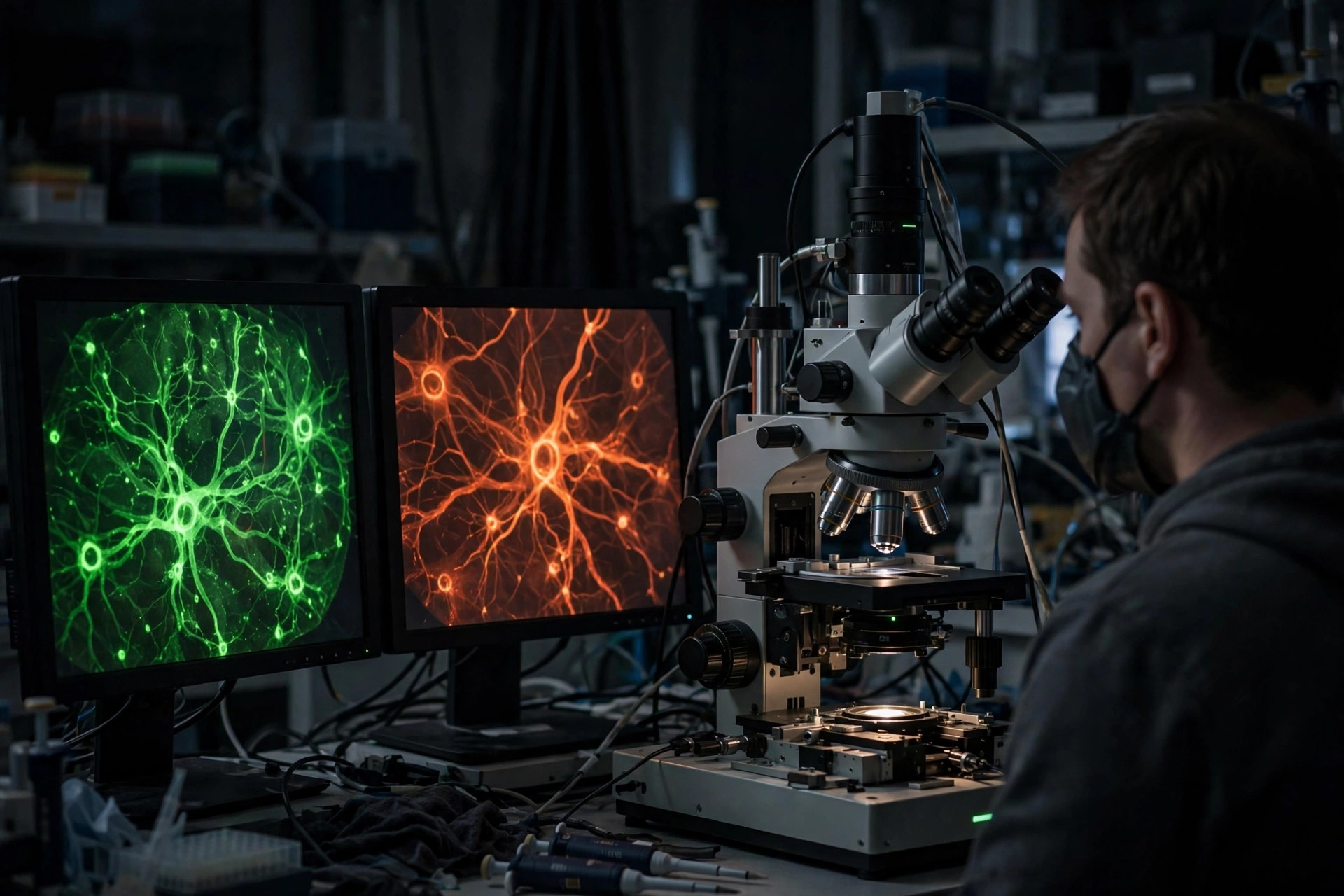
A microscope that watches brain metabolism and oxygen at the same time — in an Alzheimer's mouse model
·Tip: press play, then lock your phone or switch apps — the article keeps reading.
Two of Alzheimer's disease's recurring suspects — failing mitochondrial metabolism and impaired cerebral oxygenation — are typically investigated by different labs with different tools, which makes their relationship hard to pin down. A study in Neurophotonics (Sagar and colleagues, 2026), reported October 17 by Phys.org, combined them in a single measurement: two-photon fluorescence-lifetime and phosphorescence-lifetime imaging (FLIM-PLIM) applied to living tissue in a mouse model of Alzheimer's disease.
What does FLIM-PLIM actually measure?
Fluorescence lifetime imaging (FLIM) reads out the metabolic state of cells — including the balance of NADH, the electron carrier at the heart of mitochondrial energy production — while phosphorescence lifetime imaging (PLIM) reports local oxygen tension. Run together through a two-photon microscope, they provide metabolic and oxygen information in the same living tissue, at cellular resolution, with relatively little disruption — letting researchers watch relationships that isolated measurements cannot resolve.
Why measure metabolism and oxygen together?
Because mitochondria consume oxygen to make energy, the two variables are mechanically linked: a change in oxygen delivery alters metabolism, and a change in metabolic demand alters oxygen use. In a disease where both sides of that equation are suspected to drift, simultaneous measurement can distinguish patterns — for example, tissue that is oxygen-starved versus tissue that has oxygen but cannot use it — that point to very different underlying problems and, eventually, different interventions.
What are the honest limits?
The results come from a mouse model, so they do not establish that the same metabolic and oxygen patterns occur in people with Alzheimer's disease. This is a methods advance — an imaging framework — not a diagnostic test, a biomarker validated for clinic use, or a treatment. Its value is in the questions it now lets researchers ask in living brain tissue over the course of disease.
Where the evidence stands
- Established: Mitochondrial dysfunction and impaired cerebral oxygenation are both implicated in Alzheimer's disease and are mechanically linked through oxygen-consuming energy production.
- Preliminary: Two-photon FLIM-PLIM microscopy captured metabolic and oxygen information simultaneously in living tissue in an Alzheimer's mouse model (Neurophotonics, 2026), providing an in vivo framework for studying how the two change together.
- Absent: Evidence that the same patterns occur in human Alzheimer's disease, and any clinical diagnostic or treatment application of this imaging approach.
Join the discussion
To get the conversation started: What questions does this story raise for you? Share them below — every comment is reviewed by our moderators before it appears.
Community discussion only — not medical advice. Every comment is reviewed by our moderators before it appears. Sign in with Google or a free Hyvor account to join in.