
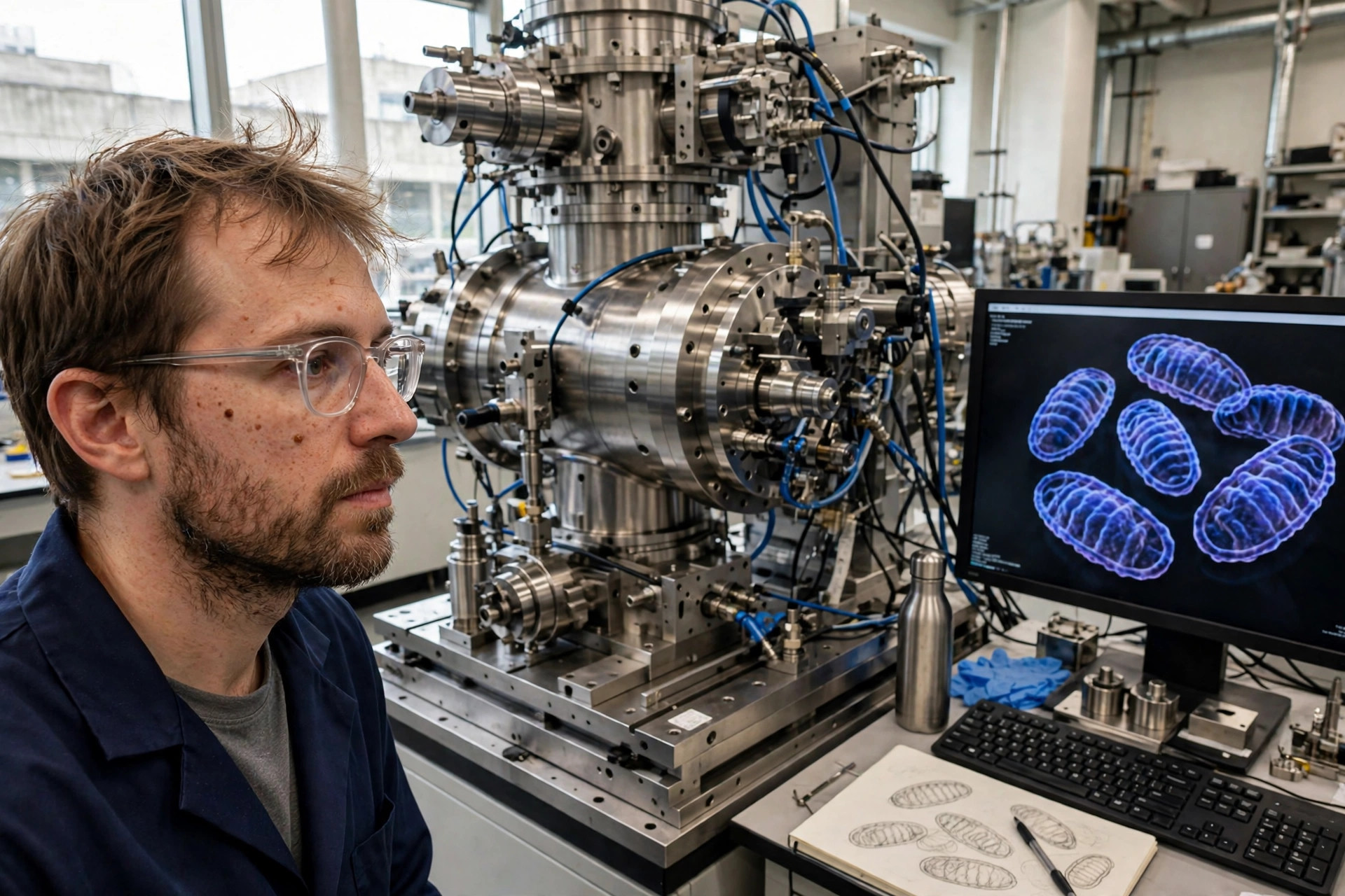
Sharper math, not sharper lenses: X-ray imaging can now pick out a single mitochondrion in 3D
Edited by Samael A. Tejada, Founder & Editor-in-Chief
·Tip: press play, then lock your phone or switch apps — the article keeps reading.
A new reconstruction method for X-ray nanotomography improved resolution about threefold, to roughly 20 nanometers, making individual mitochondria and synapses visible in three dimensions inside larger, unsliced samples (Optica, reported November 10, 2026). It is an imaging-methods advance demonstrated on research samples — not a clinical scan, and not a test patients can get.
The bottleneck was not the X-ray machine. It was the math that turns its flood of data back into a picture — and better math just made mitochondria appear.
What did the method achieve?
Using a scalable joint reconstruction approach (the bilinear Hessian method), the team extracted significantly more information from the same kind of measurement data, improving resolution around threefold to structures as small as 20 nanometers. In the study's images, an arrow picks out synapses between brain cells and another a mitochondrion inside a cell — the scale at which mitochondrial shape, position and contacts with other structures can be studied directly.
Why not just use electron microscopy?
Electron microscopy delivers extremely fine detail, but for 3D imaging the sample must be cut into very thin slices, and the process permanently damages the material. X-rays can image much larger samples in three dimensions without cutting them apart — the challenge is the enormous data volume, which is where the new mathematics and optimization algorithms come in, per Carlsson.
What does this mean for mitochondrial research?
Mitochondrial medicine increasingly turns on architecture — cristae shape, contact sites, position within the cell — and those are three-dimensional questions. A nondestructive 20-nanometer 3D method lets researchers ask them in bigger, more intact samples. It is a laboratory instrument advance, not a diagnostic: no patient imaging, no clinical validation, and no timeline to either is claimed by the authors.
Frequently asked questions
What can the new X-ray method see?
Structures down to about 20 nanometers in three dimensions — including individual mitochondria and synapses — inside samples much larger than electron microscopy can handle, without slicing them.
What was actually improved?
The mathematics of reconstruction, not the X-ray optics. The team's bilinear Hessian method extracts more information from the same measurement data, improving resolution about threefold.
Can patients get this scan?
No. It is a research imaging method demonstrated on laboratory samples, with no clinical application claimed.
Where the evidence stands
- Established: Electron microscopy gives extremely fine detail but requires thin slicing and damages samples; X-ray nanotomography can image larger samples in 3D, limited by reconstruction of very large datasets.
- Preliminary: The threefold resolution improvement to ~20 nm: one methods paper (Optica, 2026) from an international collaboration, demonstrated on research samples. Independent adoption and benchmarking are still ahead.
- Absent: Clinical evidence of any kind — this is not a patient scan or diagnostic test, and no clinical use is claimed.
Join the discussion
To get the conversation started: What questions does this story raise for you? Share them below — every comment is reviewed by our moderators before it appears.
Community discussion only — not medical advice. Every comment is reviewed by our moderators before it appears. Sign in with Google or a free Hyvor account to join in.